By Prof. Saeed Abuel Makarem
410 likes | 833 Vues
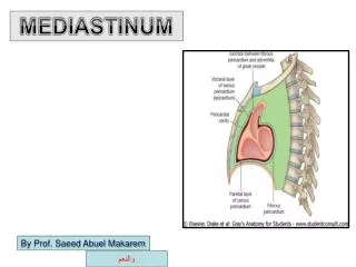
THE HEART. By Prof. Saeed Abuel Makarem. Pericardium A fibro-serous sac Surrounds the heart & proximal part of its great vessels ( Aorta , Pulmonary artery, SVC, IVC, & 4 pulmonary veins) Formed of: Outer fibrous layer Inner serous sac Serous sac has 2 layers:

By Prof. Saeed Abuel Makarem
E N D
Presentation Transcript
THE HEART By Prof. Saeed Abuel Makarem
Pericardium A fibro-serous sac Surrounds the heart & proximal part of its great vessels (Aorta, Pulmonary artery, SVC, IVC, & 4 pulmonary veins) Formed of: Outer fibrous layer Inner serous sac Serous sac has 2 layers: Parietal & Visceral (epicardium)
Fibrous Pericardium Conical or flask shape Apex: directed upwards fused with adventitia of 3 big vessels Base: rests on diaphragm Fuses with central tendon of diaphragm. Posteriorly: Separated by post. Mediasinum from middle four thoracic vertebrae (5 to 8) Anteriorly: body of sternum, costal cartilages from 2 to 6, ant. Border of lung & pleura, remains of thymus gland & two Sterno-pericardial ligaments Extends from 2nd to 6th rib
Located in the middle mediastinum • posterior to the body of sternum and 2nd-6th costal • cartilages • Anterior to T5-T8 vertebrae • 1-1.5 cm to the right of the sternum • 5-7.5 cm to the left of median plane at the level of 5th intercostal space
Function of serous pericardium: 1- Lubrication to prevent friction 2- Prevent adhesion of the heart to its surrounding
Function of fibrouspericardium 1- Maintain the central position of the heart 2-Keeps large vessels open 3-Helps venous return 4-Acts as a wall for the serous pericardium. 5- Prevents over loading & overdistention of the heart Serous pericardium Serous sac that has been invaginated by developing heart in the fetal life Visceral layer: Epicardium. Parietal layer: Lines the fibrous pericardium Pericardial cavity: potential space.
Sinuses of pericardium 2 sinuses in the serous pericardium are formed during development of the heart (Transverse & Oblique) Transverse sinus A recess behind pulmonary trunk & ascending aorta Boundaries: Ant: Pulmonary trunk & ascending aorta. Post: SVC &Upper part of the 2 atria Above: Rt. Pulmonary artery Below: the 2 atria mainly Lt.
Oblique sinus It separates the base of heart (left atrium) from posterior mediastinum (descending aorta & esophagus) Boundaries: Ant: back of Lt. atrium Post: fibrous pericardium & posterior mediastinum (esophagus & descending aorta ) Left: 2 Left pulmonary veins Right: 2 right pulmonary veins & IVC
Blood Supply Arterial Supply: Mainly supplied by pericardiophrenic and musculophrenic arteries, branches of internal thoracic Also supplied by pericardial branches of bronchial, esophageal and superior phrenic arteries Visceral layer of the serous pericardium (epicardium) supplied by the branches of the coronaryarteries Venous drainage: Veins are tributaries of azygos system. Pericardiophrenic veins also drain into the internal thoracic vein
Nerve Supply The fibrous pericardium and the parietal layer of the serous pericardium are supplied by the phrenic nerves. The visceral layer of the serous pericardium is innervated by (autonomic fibers) branches of the sympathetic trunks and the vagus nerves
Heart Muscular pump that keeps circulation going on. It is the size of hand’s fist of the same person 2/3 of its breadth lies to left of median plane and 1/3 right to median plane It is conical in shape having an apex, base, Sterno-costal, diaphragmatic, surfaces and right, and left borders.
External Features: Surfaces The heart has: Sternocostal (Anterior) surface Diaphragmatic (Inferior) surface Base (Posterior surface) Apex
Apex Directed downwards, forwards & to left Lies in LT. 5th Intercostal space 3 & ½ inches ( 9 cm) from median plane. Formed only by LT. ventricle. Pericardium & diaphragm separate the apex from fundus of stomach
Base Directed backwards and slightly to right. Formed by 2 atria mainly the left atrium. Separated from post Mediastinum by oblique sinus of pericardium Lies opposite middle 4 thoracic vertebrae (5 to 8) Bounded inferiorly by coronary groove that lodge the coronary sinus.
*Sterno-costal surface Lies behind body of sternum & 3rd to 6th costal cartilages. Divided by coronary groove into: Atrial part: Rt. Atrium Ventricular part: the two ventricles, mainly Rt. Borders: Upper: Straight, formed by the 2 atria but concealed by the ascending aorta & pulmonary trunk. Lower: Concave formed by Rt. Ventricle & small apical part of Lt ventricle Rt.: Convex formed by Rt. Atrium Lt.: Convex ,formed by Lt. auricle and Lt. ventricle.
Diaphragmatic surface Left 2/3 of this surface are formed by Lt ventricle Rt. 1/3 is formed by Rt. Ventricle The 2 ventricles are separated by post. Interventricular groove. The groove lodges the post. Interventricular artery & middle cardiac vein
Surface anatomy of Heart Point 1: Lower border of 2nd Lt costal cartilage one & half inches from the median plane. Point 2: Upper border of Rt. 3rd costal cartilage one inch from median plane. Point 3: Upper border of Rt. 6th costal cartilage one inch from its junction with the sternum. Point 4: Apex Lt. 5th Intercostal space 3 ½ inches from median plane.
Surface anatomy of the valves • Pulmonary: Left 3rdsternocostal junction • Aortic: Left 3rdIntercostal space just left to sternum • Mitral: Left 4thsternocostal junction • Tricuspid: 4thIntercostal space near the middle line.
Surface anatomy A Auscultation of the cardiac valves
Arterial Supply It is supplied by right & left coronary arteries Coronary arteries arise from the ascending aorta immediately above the aortic valves Coronary arteries and their branches are distributed over the surface of the heart lying within the subepicardial connective tissue
Right Coronary Artery Arises from anterior sinus of ascending aorta Runs between the pulmonary trunk and right auricle Runs in the atrioventricular groove At the inferior border of the heart it turns posteriorly in the atrioventricular groove Anastomosis with a branch from the left coronary artery in the posterior interventricular groove
Right Coronary Artery: Branches 1-Right conus artery 2-Anterior ventricular branches: 2-3 in number, 3-Marginal branch 4-Posterior ventricular branches, which gives a branch to atrioventricular node 6-Posterior interventricular artery 7-Atrial branches, & artery of the sinuatrial node which also supplies atria
Left Coronary Artery Larger than the right Supplies major part of the heart Arises from the left posterior aortic sinus Runs between the pulmonary trunk and left auricle Runs in the atrioventricular groove Divides into anterior interventricular & circumflex branches
Left Coronary Artery: Branches Anterior interventricular artery, which gives a small conus artery Circumflex artery, gives: Left marginal Anterior ventricular Posterior ventricular Atrial branches
Coronary Artery Anastomoses Although anastomosis do exist between the terminal branches of the right and left coronary arteries, But these are not large enough to compensate for any sudden blockage of a large branch. A sudden blockage of the larger branches results in myocardial infarction.
Venous Drainage of the Heart Most of the blood drains into the right atrium through the coronary sinus. Coronary sinus: Lies in the posterior part of the atrioventricular It is the continuation of the great cardiac vein Opens into the right atrium to the left of the inferior vena cava Tributaries: small and middle cardiac veins, posterior vein of the left ventricle, oblique vein of the left atrium Anterior cardiac veins drain directly into the right atrium Vena cordis minimi open directly into heart chambers