Download

1 / 20
200 likes | 368 Vues
Implantation. By Prof. Saeed Abuel Makarem. Implantation By. Prof. Saeed Abuel Makarem. Cleavage of Zygote. Begins about 30 hrs after fertilization Zygote divides first into 2 then 4 then 8 & 16 cells Zygote is within the thick zona pellucida during cleavage

E N D
Implantation By Prof. Saeed Abuel Makarem
Implantation • By. Prof. Saeed Abuel Makarem
Cleavage of Zygote • Begins about 30 hrs after fertilization • Zygote divides first into 2 then 4 then 8 & 16 cells • Zygote is within the thick zona pellucida during cleavage • Zygote migrates in the uterine tube from its lateral end to its medial end. • Zona pellucida is translucent under light microscope
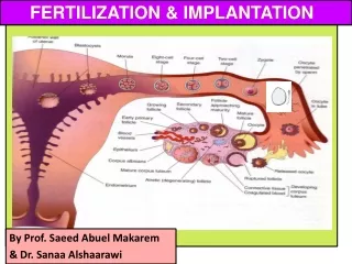
Definition of implantation: • It is the process by which the Blastocyst penetrates the superficial (Compact) layer of the endometrium of the uterus. • Site: • The normal site of implantation is the posterior wall of uterus near the fundus. • Time: • It begins about the 6th day after fertilization. • It is completed by the 11th or 12th day.
Mechanism: • The Morula reaches the uterine cavity by the 4th day after fertilization, & remains free for one or two days Fluid passes from uterine cavity to the Morula through the zona pellucida. • Now the Morula is called Blastocyst, its cavity is called blastocystic cavity, its cells divided into Embryoblast & Trophoblast.
The embryoblast projects into the blastocystic cavity, while the trophoblast forms the wall of the blastocyst. • Zona pellucida degenerates & disappears by the 5th day to allows the blastocyst to increase in size and penetrates the endometrium. By 6th day the blastocyst adheres to the endometrium • By 7th day, Trophoblastdifferentiated into 2 layers: Cytotrophblast, inner layer, mitotically active. Syncytiotrophoblast (outer multinucleated mass, with indistinct cell boundary. By 8th day the blastocyst is superficially embedded in the compact layer of the endometrium.
By the 5th day the Zona pellucida degenerates. Blastocyst begins implantation by the 6th day, (20 day of a 28 day menstrual cycle). Trophoblast cells penetrate the epithelium of the endometrium. Penetration results from proteolytic enzymes (eg.COX-2) produced by trophoblast.
Blood-filled Lacunae appear in the Syncytiotrophoblast which communicate forming a network by the day 10th or 11th • Syncytiotrophoblast erodes the endothelial lining of maternal capillaries which known as sinusoids. Now blood of maternal capillaries reaches the lacunae so Uteroplacental circulation is established by 11th or 12th day.
Endometrial cells undergo apoptosis (programmed cell death) to facilitates invasion of endometrium by the Syncytiotrophoblast. Syncytiotrophoblast engulf these degenerating cells for nutrition of the embryo. Implantation can be detected by: 1- Ultrasonography. 2- hCG (human chorionic gonadotrophin which is secreted by the Syncytiotrophoblast) about the end of 2nd week
Early Pregnancy Factor • Is an immunosuppressant protein • Secreted by trophoblastic cells • Appears in maternal serum within 24-48 hrs • It is the basis for EPT in the first 10 days of development.
By the tenth day conceptus is completely embedded in the endometrium. • For about 2 days the site of penetration shows a defect in the endometrium. • A fibrinous coagulum of blood closes this defect till the endometrial epithelium creeps over the closing plug by the 12th day to cover the defect.
Formation of embryonic disc • Embryoblast cells arranged into 2 layers: • 1- High columnar cells towards the amnion, called Ectoderm, (Epiblast). • 2- Low- cuboidal cells towards the blastocystic cavity called endoderm, (Hypoblast). • Now it is called bilaminar embryonic disc. • Formation of amniotic cavity. • A space appears between the ectoderm and the trophoblast. • Its floor is formed by the ectoderm while its roof is formed by a layer of flat cells called amniogenic cells which secretes the amniotic fluid.
Formation of Primary Chorionic villi By the 13th day Proliferation of Cytotrophblast cells produce extension inside Syncytiotrophoblast to form primary chorionic villi, Formation of the yolk sac Flat cells originate from the endoderm, form a membrane called exocoelomic membrane which lines the blastocystic cavity. Now it is called Exocoelomic cavity. The exocoelomic cavity and the exocoelomic membrane is called now primary yolk sac.
Ectopic Pregnancy: • 1- Placenta Previa. • 2- Tubal. • 3- Ovarian. • 4- Abdominal. 5- Pelvic. 6- Cervical.
Ectopic Pregnancy • It means implantation outside the uterus. • 95 to 97% of ectopic pregnancies occurs in the uterine tube. • Most are in the ampulla & isthmus. • Placenta previa: • Implantation occurs in the lower uterine segment.