Download
1 / 17
190 likes | 624 Vues
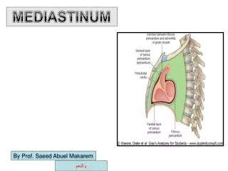
MEDIASTINUM. By Prof. Saeed Abuel Makarem. والنعم. MEDIASTINUM. DEFINTION. The median partition القسم اللوسطي (septum) of the thorax between the two pleural sacs & lungs . . Boundaries حدوده. Anterior : Sternum Posterior: Thoracic

E N D
MEDIASTINUM By Prof. Saeed Abuel Makarem والنعم
MEDIASTINUM DEFINTION The median partition القسم اللوسطي (septum) of the thorax between the two pleural sacs & lungs. Boundaries حدوده Anterior : Sternum Posterior: Thoracic vertebrae Superior: Thoracic inlet مدخل الصدر Inferior: Diaphragm On each side: pleural sacs
Divisions of the Mediastinum • For description purpose the mediastinum is divided into: • Superior علويMediastinum • Inferior سفليMediastinum • The inferior is further subdivided into: • Anterior اماميmediastinum • Middle وسطيmediastinum • Posterior خلفيmediastinum • هذه التقسيمه بواسطه الغشاء القلبي يقسم الميدياستانيوم بواسطه خط وهمي اسمه :By Transverse horizontal ويسمى :(thoracic) plane ,ويعطي القسمين اي وبي? Imaginary خياليplane passing from sternalangle التي تتوازى مع الضلع رقم 2 ( انظر الصوره )to the lower border of the body of T4 vertebrae. الخط الاحمر بالصوره
التفاصيل :اولاSuperior Mediastinum Boundaries Anterior: Manubriumsterniمقبض عظمه القص Posterior : Upper 4 thoracic vertebrae Superior : Thoracic inlet فتحه الصدر االعلويه Inferior : Transverse thoracic plane الخط الوهمي الذي سبق شرحه On each side: Pleural sacs
Contents مكوناتof Superior Mediastinum From anterior to posterior من الامام للخلفthe main contents are: • Thymus الغدة الجنينيه • Great vessels وهي: • الاورده 1- Rt& LT Brachiocephalic veins • 2- SVC • الشرايينArch of aorta & its 3 branches: • Left subclavian artery • Left common carotid artery Brachiocephalic trunk • Vagus nerve • phrenic nerve • Cardiac plexus ظفيرهof nerves • Left recurrent laryngeal nerve • Trachea • Esophagus • Thoracic duct • Prevertebral muscles
هذه الصوره مهمه جدا وتلخص الدرس ومهمه في العملي
:ثانيا1-Anterior Mediastinum A very narrow space in front of the pericardium overlapped متداخلby the anterior borders of both lungs Boundaries Anterior: Body of the sternum Posterior: Pericardium & heart Superior: Transverse thoracic plane Inferior: Diaphragm superior aspect On each side: Mediastinal pleurae
Contents of Anterior Mediastinum • Thymus الجزء السفلي منها • Sterno-percardial ligaments • Branches of the internal thoracic vessels • Fat • Lymph vessels & nodes
2- Middle Mediastinum Occupied by the pericardium & its contents along with phrenic nerves & pericardiophrenic vessels Contents Heart Ascending aorta, Pulmonary trunk, Pulmonary arteries SVC Azygos vein, Pulmonary veins (4) Phrenic nerve Deep cardiac plexus Lymph nodes Bifurcation تشعبof trachea
3- Posterior Mediastinum • Lies between: • The pericardium anteriorly • and • The vertebral column Posteriorly
Boundaries Posterior Mediastinum Anterior: Pericardium & bifurcation of the trachea Posterior: Lower 8 thoracic vertebrae On each side: Mediastinal pleura Inferior: Diaphragm Contents • Esophagus • Descending thoracic aorta and its branches • Azygos, Hemiazygos veins • Vagus nerve & splanchnic nerves • Thoracic duct
Structures seen in the Mediastinum تلخص الدرس ومهمه بالعملي Right side Left side
Esophagus • About 25 cm long tub. • Continuous above with pharynx opposite مقابلC6 vertebra • Passes through the diaphragm at the level of T10, to join the stomach • In thorax, passes downward & to the left through superior and posterior mediastinum • At the level of sternal angle, pushed to the midline by the arch of aorta
Sympathetic Trunk عصب يمتد من المخ لجميع الجسم وهو يشبه السبحه(Thoracic Part) • Continuous above with the cervical and below with the lumbar parts • Runs downward on the front of the heads of the ribs • Leaves the thorax at the side of the body of T12 vertebra by passing behind the medial arcuateligaments التي تربط الكرست – راجع درس عضلات التنفس • Has 12 segmentally arranged ganglia, each with white and grey rami communicntespassing to the corresponding spinal nerves • The first ganglion is often fused with the inferior cervical ganglion to form the stellate ganglion
Sympathetic Trunk Branches • Grey ramicommunicantes(carry postganglionic fibers) to all thoracic spinal nerves • First five ganglia give postganglionic fibers to heart, lungs, esophagus and aorta • Lower eight ganglia give passage to preganglionicfibers which form three sets of splanchnic nerves that supply the abdominal viscera • Greater splanchnic from 5-9thganglia • Lesser splanchnic from 10th &11th • ganglia • Lowest splanchnicfrom 12thganglion
MEDIASTINOSCOPY جهاز يستخدم لفحص منطقة الميدياستيرنيوم Mediastinoscopy: a diagnostic procedure اجراء for exploration of the superior mediastinum and for obtaining الحصول على specimens عينهfrom the tracheobronchial lymph nodes. The mediastinoscope is inserted through a small incision just above the suprasternal notch and can penetrate down to the bifurcation of the trachea. Used to determine the spread of carcinoma السرطان عفانا اللهof the bronchus.