Download
1 / 36
410 likes | 798 Vues
The Role of Medical Imaging Informatics in Healthcare. Outline • Medical Imaging and System Integration • Medical Imaging Informatics and CAD • Integration of CAD to PACS Operation • Image-assisted Treatment • The Creation of a Continuum – Across the Chasm from Diagnosis to Treatment.

E N D
Outline • Medical Imaging and System Integration • Medical Imaging Informatics and CAD • Integration of CAD to PACS Operation • Image-assisted Treatment • The Creation of a Continuum – Across the Chasm from Diagnosis to Treatment
Academic Excellence The Nobel Prize • 1979 “For the Development of computer assisted tomography (CAT)” Hounsfield Cormack • 2003 “For the Discoveries concerning magnetic resonance imaging (MRI)” Paul Lauterbur Peter Mansfield
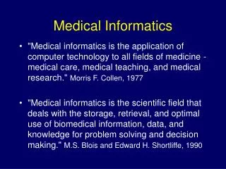
Magnetic resonance imaging (MRI), Magnetic resonance imaging (MRI), is a non-invasive method used to render images of the inside of an object. It is primarily used in medical imaging to demonstrate pathological or other physiological alterations of living tissues. Definition: Pathology is the study and diagnosis of disease through examination of organs, tissues, cells and bodily fluids Definition: Physiology is the study of the mechanical, physical, and biochemical functions of living organisms. MRI Hand animation scan MRI vs CT A computed tomography (CT) , originally known as computed axial tomography (CAT) scanner uses X-rays, a type of ionizing radiation, to acquire its images, making it a good tool for examining tissue composed of elements of a relatively higher atomic number than the tissue surrounding them, such as bone and calcifications (calcium based) within the body (carbon based flesh), or of structures (vessels, bowel) which have been artificially enhanced with contrast agents containing elements of a higher atomic number than the surrounding flesh (iodine, barium). MRI, on the other hand, uses non-ionizing radio frequency (RF) signals to acquire its images and is best suited for non-calcified tissue.
Magnetic resonance imaging (MRI) (continued) • The magnet is the largest and most expensive component of the scanner, and the remainder of the scanner is built around the magnet. Just as important as the strength of the main magnet is its precision. The straightness of flux lines within the centre or, as it is known as, the iso-centre of the magnet, need to be almost perfect. • Magnetic gradients are generated by three orthogonal coils, oriented in the x, y and z directions of the scanner. These are usually resistive electromagnets powered by sophisticated amplifiers which permit rapid and precise adjustments to their field strength and direction. • In 1983 Ljunggren[9] and Tweig[10] independently introduced the k-space formalism, a technique that proved invaluable in unifying different MR imaging techniques. They showed that the demodulated MR signal S(t) generated by freely precessing nuclear spins in the presence of a linear magnetic field gradient G equals the Fourier transform of the effective spin density i.e. MRI Animation 1
PET Positron Emission Tomography • Positron emission tomography (PET) is a nuclear medicine medical imaging technique which produces a three-dimensional image or map of functional processes or Metabolic Activities in the body. • To conduct the scan, a short-lived radioactive tracer isotope, which decays by emitting a positron, which also has been chemically incorporated into a metabolically active molecule, is injected into the living subject (usually into blood circulation). • The data set collected in PET is much poorer than CT, so reconstruction techniques are more difficult (see section below on image reconstruction of PET). PET Animation Pet Animation Atoms
Computed tomography (CT), originally known as computed axial tomography (CAT or CT scan) and body section roentgenography, is a medical imaging method employing tomography where digital geometry processing is used to generate a three-dimensional image of the internals of an object from a large series of two-dimensional X-ray images taken around a single axis of rotation
Medical Imaging • Body Region, Organ, Tissue, Cell, Gene • Diseases – What you want to detect, to see, or to diagnosis? • Energy sources • Detectors • Image formation • Display • User Interface • Connection to other Systems
Anatomy to Physiology • • Anatomy: Body regions, organs, blood • vessels, etc. • Can we see smaller anatomy? How small is • small? • Static vs. Dynamics: how fast can we detect • and record? • • Physiology: Functions, metabolism, oxygen • concentration, blood flow, etc. • How fast can we detect and record? • • Can we combine anatomy and physiology? • Can we see the dynamic?
Medical Images • One-dimensional Signals • Two-dimensional Images • Three-Dimensional Images • Four-Dimensional Images • Five- or Higher-Dimensional ? Rotating Tesseract The four-dimensional equivalent of a cube. The fourth dimension and orthogonality A right angle is defined as one quarter of a revolution. Cartesian geometry arbitrarily chooses orthogonal directions through space, which means that they are at right angles to one another. The orthogonal directions of three-dimensional space are known as the length, width and height. The fourth dimension is therefore the direction in space that is at right angles to these three observable directions.
One 256-Slice CT Scan 256 x 0.5 MB = 178 MB
Medical Imaging • Body Region, Organ, Tissue, Cell • Diseases – What you want to detect to see, or diagnosis? • Energy sources • Detectors • Image formation • Display • User Interface • Connection to another Systems
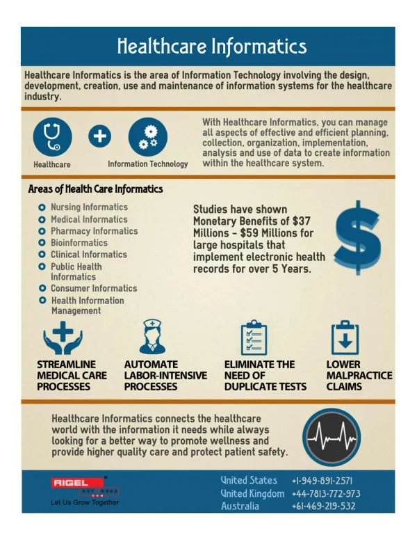
Imaging Informatics • Computer Software Technology • Mathematical Modeling • Patient Information & History • PACS and other medical Image Data • Infrastructure – Networking – knowledge base – Visualization and Presentation
One Approach to Image Storage Picture Archiving and Communication System –( PACS)
CAD (Computer Aided Diagnosis/ (Detection)) • CADx – CA Diagnosis • Provide computer output to assist human (radiologist) in image interpretation • One of the major research subjects in medical imaging and diagnostic radiology during the past 5 years • Being applied in clinical practice
Effect of CAD on Ambulatory Care and Emergency Physicians • The automated assist and diagnosis software is approaching the quality of human experts that evaluate images
The unaided scan on left was enhanced and evaluated using the CAD-diagnosis software and the picture on the right color coded the abnormality and displayed a message.
CAD-PACS (Computer Aided Diagnostics – Picture Archiving and Communications System) Integration Using DICOM & IHE (Data Integration and Communications)
Combined Diagnosis with Treatment
Ultrasound-guided Endoscopy: Imagine • Traditional endoscopy: Use fiber optic with visible light • Ultrasound endoscopy: Small transducer at the end of endoscopic tube • Two possible types of images: Circumferential image of lumen Planar image of lumen • Source: Barret’s Foundation
Ultrasound-guided Endoscopy: Example • Ultrasound endoscopy for upper GI tract • Two transducers at tip of endoscope: Low frequency: Imaging • High frequency:treatment • Source: Penn Center
Ultrasound-guided Endoscopy: Treatment • Tumor detection with ultrasound • Heating and killing tumor cells with high intensity focused ultrasound (HIFU) • Effective hyperthermia: above 70°C Mechanical result: tissue disruption • Focus: possible single transducer to send both imaging and therapeutic frequencies