Download
1 / 36
430 likes | 1.17k Vues
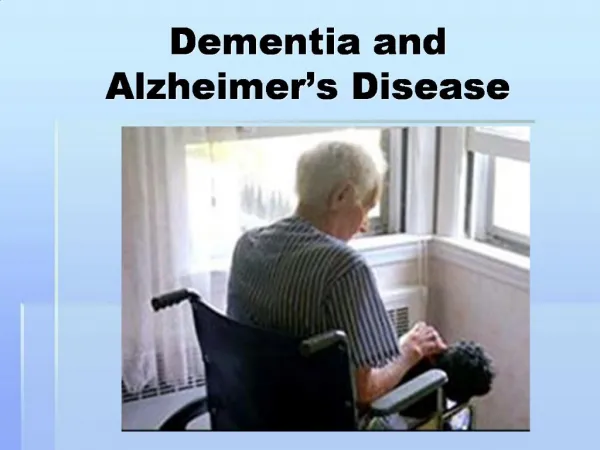
Diagnostic Assessment of Dementia and Alzheimer’s Disease: GUIDELINES. צוות הועדה יו"ר: דר' גרי סינוף יו"ר: דר' אליסה אש דר' רחל בן חיון דר' ירחמיאל ברבר דר' שלומית בר זמר מגן פרופ' יצהל ברנר דר' אלי ורטמן . אבחון קליני של ירידה קוגניטיבית ושיטיון.

E N D
Diagnostic Assessment of Dementia and Alzheimer’s Disease: GUIDELINES צוות הועדה יו"ר: דר' גרי סינוף יו"ר: דר' אליסה אש דר' רחל בן חיון דר' ירחמיאל ברבר דר' שלומית בר זמר מגן פרופ' יצהל ברנר דר' אלי ורטמן
אבחון קליני של ירידה קוגניטיבית ושיטיון • אבחנה קלינית של ירידה קוגניטיבית ובעיקר שטיון הינה חשובה על מנת לאפשר את הטיפול והתמיכה. • שיטיון אינה מאבחן את התהליך הפתופיזיולוגי ולא סיבת האטיולוגיה. • אבחון שיטיון דורש התייחסות לשני הנושאים ביחד ובנפרד.
עדכונים בתחום בהעשור האחרון • הפתולוגיה המוחית בדמנציה מתפתחת שנים לפני ההסתמנות הקלינית. • שיטיון אינם מסתמנות תמיד בליקויי זיכרון. • ההיסטופתולוגיה בספקטרום נרחב של תסמונות קליניות. • לא ניתן להבדיל בוודאות בין התסמונות השונות על פי ההסתמנות הקלינית. • סמנים ביולוגיים והדמתים בקורלציה לשינויים הפתופיזיולוגיים ותומכים בסימנים הקליניים ומאפשרים אבחנה מדויקת יותר בעוד החולה בחיים. • נמצאו מוטציות גנטיות אחראיות לתסמונות הקלינית גורמות להסתמנות מ"א בגיל צעיר והמהווה גורם סיכון למ"א הפורצת בגיל המבוגר. • האמצעים לאבחן שטיון מגוונים ומוכתבים על ידי המשאבים העומדים לרשות הקלינאים. • חוקרים מתמקדים בשימוש בסמנים קליניים, מבחנים נוירופסיכולוגים, בהדמיה, סמנים ביולוגים בנוזל השדרה או בדם, וסמנים גנטיים.
מקורות מידע • Hort J, et al., EFNS guidelines for the diagnosis and management of Alzheimer's disease. Eur J Neurol. 2010 Oct;17(10):1236-48. • McKhann GM, et al., The diagnosis of dementia due to Alzheimer's disease: recommendations from the National Institute on Aging-Alzheimer's Association workgroups on diagnostic guidelines for Alzheimer's disease. Alzheimers Dementia. 2011 May;7(3):263-9. • Albert MS, et al.;The diagnosis of mild cognitive impairment due to Alzheimer's disease: recommendations from the National Institute on Aging-Alzheimer's Association workgroups on diagnostic guidelines for Alzheimer's disease. Alzheimers Dementia. 2011;7(3):270-9. • Other sources: • Tartaglia MC, Rosen HJ, Miller BL. Neuroimaging in dementia. Neurotherapeutics. 2011 8(1):82-92. • Blennow K, Zetterberg H. Cerebrospinal fluid biomarkers for Alzheimer's disease. J Alzheimers Dis. 2009;18(2):413-7. • Goldman JS, et al.; Genetic counseling and testing for Alzheimer disease: joint practice guidelines of the American College of Medical Genetics and the National Society of Genetic Counselors.Genet Med. 2011 Jun;13(6):597-605.
אבחון קליני • קבלת היסטוריה ובדיקה גופנית. • ביצוע אבחון קוגניטיבי. • הערכה תפקודית והתנהגותית. • בירור רפואי לצורך אבחון מחלות נילוות ושיפור דיוק האבחנה.
אנמנזה רפואית • נתונים דמוגרפיים ובריאותיים. • תאור תהליך הירידה בקוגניציה. • מידע על תפקוד, בסיסי ואינסטרומנטלי. • רישום מפורט על טיפול תרופתי. • אנמנזה בבעיות נפשיות ובשינה. • הערכה התנהגותית. • מספר ביקורי מרפאה לקבוע את האבחנה נע בין 1-3.
הרכב צוות הרכב הצוות תלוי במשאבים העומדים לרשות המרפאה. מומלץ על צוות רב מקצועי: רופא מומחים בשיטיון גריאטריה נוירולוגיה פסיכיאטריה מאבחנת לפרש מצב קוגניטיבי אחיות לנטר את התוכניות הטיפולית ולתת הדרכה למטופלים ולבני משפחה פיזיותרפיסט ומרפא בעסוק לשמירת תפקוד עובד סוציאלי לבנות תוכנית
הערכה תפקודית, נפשית והתנהגותית • נדרשת התייחסות לתפקודבסיסי ואינסטרומנטלי • תפקוד בסיסי כולל נטילת תרופות • ניהול חשבון בנק ותשלום חשבונות • שימוש במכשירים או יכולת להשתלט על מכשיר חדש • ביצוע קניות, נהיגה ושימוש בתחבורה ציבורית • כלים ולידים להערכת תפקוד אינסטרומנטלי • Blessed Functional Activity Scale • Lawton and Brody IADL scale • Modified Barthel Index • Functional Activities Questionnaire
בדיקה גופנית • בדיקה גופנית לאבחון שיטיון חייבת לכלול בדיקה נוירולוגית מקיפה עם דגש על: • המערכת האקסטראפירמידלית • המערכת המוחון • בדיקת הליכה והחזרי שיווי משקל • בדיקת תפקודים פרונטלים כולל סימני שיחרור פרונטלים • סימנים אחרים של הפרעה בתפקוד קורטיקלי (הפרעה תחושה פריאטלית, פרקסיס)
אמצעי אבחון במבחנים נוירופסיכולוגים מבחני סינון MMSE: בודק מספר תחומים קוגניטיביים ואורך בין 10-15 דקות. שימושו העיקרי במרפאות הקהילה הוא לשלול דמנציה נועד לקבוע את חומרת הדמנציה מבחן ציור שעון: בשימוש באופן נרחב ככלי סריקה לדמנציה. תלוי פחות מה MMSE בשפה והשכלה MoCA /STMS/SLUMS: פותחו ככלי סריקה ל-MCI ולדמנציה. מבחנים נוירופסיכולוגים פסיכומטרים מבחני זיכרון וזיכרון אפיזודי מבחני תחומים נוספים תפקודים ניהוליים תפקודי שפה תפקודי תפיסה מרחבית (כולל זיכרון וויזואלי) מבחן להעריך שיפוט
אמצעי אבחון במבחנים נוירופסיכולוגים: המלצות • מומלץ לבצע מבחן סינון בכל מקרה שיש לנבדק תלונות קוגניטיביות, דיווח ממוסר מידע, או חשד לקליני לירידה • סוג המבחן תלוי בחומרת המצב הקליני של הנבדק המתברר מהערכה הקלינית של מומחה: • מומלץ על ביצוע MMSE וציור שעון בנבדקים עם ירידה קוגניטיבית בולטת. • מומלץ על ביצוע MOCA בנבדקים בלי סיפור של ירידה תפקודית משמעותית • MMSE תקין לא שולל שיטיון או MCI: מומלץ להוסיף בדיקה רגישה יותר לפי החשד הקליני. • ידוע על איתור חיובי שגוי (False Positive) ואיתור שלילי שגוי (False Negative). • מומלץ על ביצוע אבחון נוירופסיכולוגי שיש התלבטות אבחנתית לגבי האם קיימת הפרעה או סוג של הפרעה, או שחשוב לפרט את הקף הפגיעה
בדיקות עזר לזיהוי מצבים הפיכים וזיהוי אפשרות של אבחנה חלופית בדיקות שגרתיות בדיקות הדמיה מבנית בדיקות הדמיה תפקודית סמנים ביולוגיים בדיקות גנטיות
הגורמים שרצוי לשלול כגורם אפשרי לירידה קוגניטיבית לשלול מחלה רפואית חריפה לשלול מחלת נפש בעיקר דיכאון
בדיקות מעבדה • מומלץ לשלול תהליכים נילווים עלולים להשפיע על תפקוד קוגניטיבי • בדיקות דם: ס"ד, כימיה מלאה כולל סידן, תפקוד בלוטת התריס, ויטמין 12B, חומצה פולית • בדיקות נוספות על פי ההיסטוריה הרפואית/בדיקה גופנית/תוצאות בדיקות המעבדה: • שקיעת דם, בירור ראומטולוגי, סרולוגיות למחלות זיהומיות או דלקתיות, מדדים של גורמי סיכון ווסקולרים (שומנים, המוגלובין מסוכר), ועוד.
המקום של בדיקות הדמיה באבחון שיטיון הדמיה מוחית מבנית CT מוח בלי חומר ניגוד: מעודפת לשם אבחנה של דימום, וגם ניתן לוודא מקרים של אטרופיה מוחית מתקדם. MRI מוח: בדיקה מעודפת לזיהוי שינוים מבנים קורטיקלים ותת-קורטיקלים, הפרעה בגזע המוח, ושינויים איסכמיים הקשורים למחלה נוירו-וסקולרית, או בזיהוי פתולוגיות נלוות אחרות. בדמנציות אחרות, MRI רגיש יותר מ-CT לזהות סימנים של אטרופיה מוקדית מומלץ על ביצוע הדמית מוח מבנית באבחון שיטיון מומלץ על ביצוע CT כבדיקה ראשונה מומלץ על MRI מוח בחשד לתהליך איסכמי או התלבטות אבחנתית מומלץ MRI כבדיקה הראשונה עם חשד לתהליך נאופלסטי או דלקתי
הדמית מוח תפקודית • קיימות שיטות של הדמית מוח תפקודית היעילות באבחנת דמנציה: • SPECTSingle Positron Emission Computed Tomography • FDG-PET Fluorodeoxyglucose Positron Emission Tomography • SPECT: יעילות קלינית מתונה לאבחן מ"א, MCI, דמנציות אחרות • :FDG-PET בעל רגישות ורצולוציה יותר גבוהות מ-SPECT, יקר ופחות זמין • יש לבצע הדמייה תפקודית בלאבחן בין מ"א לבין דמנציות אחרות במידה והאבחנה בהתלבטות(British National Institute for Health and Clinical Excellence, 2006) • לא מומלץ על ביצוע הדמיה תפקודית באופן רוטיני עקב השפעתם של תהליכים נילווים כולל הפרעות נפשיות על תפקוד המוח וקצב איתור חיובי שגוימשמעותי של הדמייה לקויה במקרים שאין מחלת שיטיון כאבחנה בסיסית. • ניתן לשקול ביצוע במקרים עם התלבטות קלינית לגבי האבחנה הבסיסית
ביצוע ניקור מותני כחלק מאבחון שיטיון הוא חלק חשוב במקרים של הפרעות קוגניטיביות המופיעות יחסית מהיר (Rapidly Progressive Dementia) ניקור מותני למדוד את חלבוני TAU או 14-3-3 יכול להיות בדיקה יעילה מאוד באבחנת מחלת פריונים (CJD) ניתן לזהות סמנים ביולוגיים למ"א בנוזל השדרה (CSF) עמילואיד- ביתא ( ( beta-amyloid 1-42 סך חלבון טאו (T-tau) תת סוג של טאו (Phosphorylated Tau ,p-tau) יחס Aβ42/tau ב-CSF למרות רגישות סבירה לגלות מ"א, יש עדיין בעיות בספציפיות לאבחן בין סוגים אחרים של שטיון, בעיות באחידות ביצוע במעבדות שונות. לא מומלץ על ביצוע ניקור מותני לזהות סמנים ביולוגים כחלק מהאבחון הרוטיני בשלב זה, רק במקרים מיוחדים כשהתמונה לא טיפוסית כמו אנשים צעירים מגיל 55-60 או לאנשים עם דמנציה המתקדמת במהירות לשלול סיבות אחרות של ירידה קוגניטיבית (RAPIDLY PROGRESSIVE DEMENTIA).
אמצעי אבחון גנטי • חלק קטן של שיטיון מוסבר ע"י מוטציות גנטיות. • בכל המחלות הנוירודגנרטיביות יש השפעה של גנים שונים, ומוטציות מתוארות במחלות שונות. • (AD) AUTOSOMAL DOMINANT • APP, 1PSEN , 2PSEN . • כ-5% ממוטציות, רובם אצל אנשים צעירים מתחת לגיל 60. • אין המלצה חד משמעית לאנשים צעירים סמפטומטים עם רקע משפחתי. • מומלץ בדיון לפני ביצועה ולהפנות לייעוץ גנטי. • אין המלצה חד משמעית בקרובי משפחה של נבדק בעל מוטציה. • במדינות אחרות פועלים לפי נוהלי בדיקה גנטית דומים למחלת הנטינגטון • לא מומלץ על ביצוע בדיקה גנטית אצל ילדים • APOE-SUSCEPTIBILITY GENE • המעלה רגישות לחלות עם מ"א בקרב אנשים בעלי פולימורפיזם APOEe4 • למרות הקשר, רוב המקרים לא מוסברים ע"י פולימורפיזם זה • בדיקה גנטית ל-APOE מבוצע אך ורק במסגרת מחקרית. • כיום אין המלצה על בדיקות גנטיות במסגרת בירור של דמנציה בגיל קשיש.
הקריטריונים לאבחון שיטיון McKhann 2011 • שיטיון היא אבחנה קלינית הקשור במצב תפקודי. • חייבת להיות ירידה בתפקוד קוגניטיבי המפריעה בתפקוד בפעילויות רגילות ומשקף ירידה מרמות קודמות של תפקוד וביצוע. • ניתן לאחבן מצב של שיטיון במידה ויש תסמינים קוגניטיבים, התנהגותיים או נוירופסיכיאטרים הגורמים להפרעה בתפקוד בעבודה או בפעילויות שיגרתיות. • המצב לא מוסבר ע"י דליריום או הפרעה פסיכיאטרית מג'ורית. • הפרעה קוגניטיבית או התנהגותית אמורה לערב לפחות שני תחומים של חשיבה כולל: זיכרון, שיפוט לקוי, הסקה לקויה, הפרעה בביצוע מטלות מורכבות, הפרעה בתפיסה מרחבית, תפקודי שפה לקויים, או שינויי אישיות והתנהגות. • הליקוי הקוגניטיבי יזוהה באמצעות שילוב נתונים מההיסטוריה הרפואית, הבדיקה הקלינית וההערכה הקוגניטיבית. • במקרים בהם לא ניתן להגיע למסקנה ברורה יש להפנות את הנבדק להערכה נוירוקוגניטיבית במסגרת מרפאה מקצועית.
קריטריונים אבחנתיים של מ"א • קריטריונים מבוססים על הסכמה של דעות קליניות של מומחים בתחום. • קריטריונים לאבחנה קלינית של מ"א נקבעו ע"י קבוצת עבודה של NINCDS-ADRDA ב-1984 • לאחרונה ע"י קבוצת עבודה המכון הלאומי לזקנה של ארה"ב ועמותת אלצהיימר אמריקאית (NATIONAL INSTITUTE ON AGING-AA) שפורסמו 2011.
שיטיון ע"י מ"א בסבירות גבוהה (PROBABLE AD DEMENTIA) • הנבדק עומד בקריטריונים של שיטיון ובעל כל המאפיינים הבאים: • התחלה והתפתחות הדרגתית משך חודשים עד שנים • הפרעה מתקדמת והחמרה בתפקודים קוגניטיבים ע"י דיווח או תצפית • הסמנים הקוגניטיבים הראשונים והבולטים הם מסוג של • הצגה אמנסטית (מצב הטיפוסי) או • הצגה נונ-אמנסטית (הפרעה שפתית, דיסאקסקיוטיבית, תפיסה מרחבית) • אבחנה זאות לא ניתן במידה שיש עדות ל: • מחלה צרברווסקולרית מלווה ניכרת, • מאפיינים בסיסים של שיטיון ניווני אחר, או • עדות לתהליך נוירולוגי פעיל אחר או מחלה אחרת העלולה להסביר את המצב או שימוש תכשיר תרופתי העלול להפריע בקוגניציה בצורה משמעותית. • לציין שכל נבדקים בעלי קריטריונים למ"א בסבירות גבוהה (PROBABLE AD DEMENTIA) לפי הקריטריונים המעודכנים גם היו מאופיינים כחולי מ"א בסבירות גבוהה לפי הקריטריונים הישנים של NINCDS-ADRDA
שיטיון ע"י מ"א בסבירות בינונית (Possible AD Dementia) • בעל כל קריטריוני אבחנה של שיטיון בסבירות גבוהה (PROBABLE AD DEMENTIA) אבל: • ידוע על מהלך אטיפי מסוג של תחילה פתאומית או • שאין מספיק מידע אנמנזה או מידע אובייקטיבי לתעד מהלך פרוגרסיבי. • ממלא כל הקריטריונים לשיטיון בסבירות גבוהה (PROBABLE AD DEMENTIA) אבל: • יש עדות לתהליך פתולוגי נלווה אחר, מסוג של • (1) מחלה צרברווסקולרית מלווה ניכרת, • (2) נוכחותם של מאפיינים בסיסים של שיטיון עם גופיפי לווי, או • (3) עדות לתהליך נוירולוגי פעיל אחר או מחלה אחרת העלולה להסביר את המצב או שימוש תכשיר תרופתי העלול להפריע בקוגניציה בצורה משמעותית. • לא כל נבדק בעל קריטריונים לשיטיון ע"י מ"א בסבירות בינונית (POSSIBLE AD DEMENTIA) לפי קריטריוני NINCDS-ADRDAהיה עומד בקריטריונים המעודכנים. • למשל, ניתן לאבחן מ"א בסבירות בינונית (POSSIBLE AD DEMENTIA) במקרים של חשד לשני סוגי פתולוגיה בו זמניים.
לקוי קוגניטיבי מתון (MCI) נבדק שמהווה ירידה קוגניטיבית אך אין מצב של שיטיון. הקריטריונים הגרעינים לאבחנה זו הם: דאגה על ירידה קוגניטיבית מהנבדק, ממוסר מידע ידיעני, או מקלינאי מיומן המכיר טוב את הנבדק עדות לליקויים בתחום אחד או יותר של קוגניציה עדות לביצוע ירוד בתחום קוגניטיבי אחד או יותר ממה צפוי רקע הנבדק (כולל גיל ורמת השכלה) שמירה של עצמאות ביכולת וביצוע תפקודי למרות שינויים קלים בתפקוד אינסטרומנטלי, נבדק מחזיק את עצמאו עם עזרה מינימלית. לא קיים מצב של שיטיון MCI אמנסתי (AMNESTIC-MCI) ע"י ליקויים אוביקטיבים בזיכרון אפיסודי, MCI עם ליקוי קוגניטיבי בלי ליקויים בתחום הזיכרון (NONAMNESTIC-MCI). SINGLE OR MULTIPLE DOMAIN MCI עדות אוביקטיבית לירידה קוגניטיבית קוי יסוד של ציונים במבחנים פסיכומטרים לפחות כ1-1.5 סתיות תקן מתחת לממוצע בהשוואה לאכלוסיה של אנשים תואמי גיל ורמת השכלה עדות להחמרה פרוגרסיבית והתקדמות בליקויים הקוגניטיבים עם זמן הערכות קליניות וקוגניטיבית חוזרות כדי להעיד על דיוק האבחנה ולהערכה של התערבויות טיפוליות. מומלץ על שימוש בבדיקות מספיק רגישות לזיהוי הפרעות קלות יותר. אבחון נוירופסיכולוגי פסיכומטרי במידה והאבחנה לא מספיק ברורה
דמנציה פרונטו-טמפורלית • Neary et al., 1998 • Insidious onset and gradual progression • Early decline in social interpersonal conduct • Early impairment in regulation of personal conduct • Early emotional blunting • Early loss of insight
FTD Criteria Consortium Revised criteria for bvFTD 2010 • Possible bvFTD • Progressive deterioration of behavior or cognition with early appearance (first 3 years of sx) of at least 3 of the following characteristics: • behavioral disinhibition • apathy or inertia • loss of sympathy or empathy • Perseverative, stereotyped, or compulsive/ritualistic behavior • Hyperorality or dietary changes • Neuropsychological profile: executive/generation deficits with relative sparing of memory and visuospatial functions • Probable bvFTD: Clinical syndrome with supportive imaging • bvFTD with definite FTLD pathology • Exclusion criteria • Pattern of deficits is better accounted for by other non-degenerative nervous system or medical disorders or psychiatric disorder • Presence of biomarkers consistent with AD
Primary Progressive Aphasia (PPA) • Inclusion Criteria • Most prominent clinical feature is difficulty with language: e.g., word-finding deficits, paraphasias, effortful speech, grammatical and/or comprehension deficits • These deficits are the principal cause of impaired daily living activities: e.g., problems with communication activity related to speech and language, such as using the telephone; or performing routine job responsibilities that require verbal communication • Aphasia should be the most prominent deficit at symptom onset and for the initial phases of the disease. • Exclusion Criteria • Pattern of deficits is better accounted for by other non-degenerative nervous system or medical disorders: e.g., neoplasm, cerebrovascular disease, hypothyroidism • Cognitive disturbance is better accounted for by a psychiatric diagnosis: e.g., depression, bipolar disorder, schizophrenia, pre-existing personality disorder • Prominent initial episodic memory, visual memory and visuo-perceptual impairments: e.g., inability to copy simple line drawings • Prominent initial behavioral disturbance: e.g., marked disinhibition, emotional detachment, hyperorality or repetitive/compulsive behaviors
Semantic Variant PPA (SV-PPA) • Core criteria • Poor confrontation naming (of pictures or objects), particularly for low familiarity or low frequency items • Impaired single-word comprehension • Supportive criteria (at least 3) • Poor object knowledge, particularly for low frequency or low familiarity items • Surface dyslexia and/or dysgraphia • Spared repetition • Spared motor speech (no distortions) and grammar • Imaging-Supported SV-PPA Diagnosis • 1. Clinical diagnosis of SV-PPA • 2. Imaging must show one or more of the following results: • 1. Predominant anterior temporal lobe atrophy on MRI • 2. Predominant anterior temporal hypoperfusion or hypometabolism on SPECT or PET
Nonfluent/Agrammatic Variant PPA (NFAV-PPA) • Core criteria • 1. Agrammatism in language production • 2. Effortful, halting speech with inconsistent distortions, deletions, substitutions, insertions, or transpositions of speech sounds, particularly in polysyllabic words (often considered to reflect "apraxia of speech") • Supportive criteria (at least 2) • 3. Impaired comprehension of syntactically complex sentences • 4. Spared single word comprehension • 5. Spared object knowledge • Imaging-Supported NFAV-PPA Diagnosis • 1. Clinical diagnosis of NFAV-PPA • 2. Imaging must show one or more of the following results: • 1. Predominant left posterior fronto-insular atrophy on MRI • 2. Predominant left posterior fronto-insular hypoperfusion or hypometabolism on SPECT or PET
דמנציה עם גופיפי לווי (DLB)McKeith et al., 2005 • 1. Central feature (essential for a diagnosis of possible or probable DLB) • Dementia defined as progressive cognitive decline of sufficient magnitude to interfere with normal social or occupational function.Prominent or persistent memory impairment may not necessarily occur in the early stages but is usually evident with progression. Deficits on tests of attention, executive function, and visuospatial ability may be especially prominent. • 2. Core features (two core features are sufficient for a diagnosis of probable DLB, one for possible DLB) • Fluctuating cognition with pronounced variations in attention and alertness • Recurrent visual hallucinations that are typically well formed and detailed • Spontaneous features of parkinsonism • 3. Suggestive features (If one or more of these is present in the presence of one or more core features, a diagnosis of probable DLB can be made. In the absence of any core features, one or more suggestive features is sufficient for possible DLB. Probable DLB should not be diagnosed on the basis of suggestive features alone.) • REM sleep behavior disorder • Severe neuroleptic sensitivity • Low dopamine transporter uptake in basal ganglia demonstrated by SPECT or PET imaging
דמנציה עם גופיפי לווי (DLB)McKeith et al., 2005 • 4. Supportive features • Repeated falls and syncope • Transient, unexplained loss of consciousness • Severe autonomic dysfunction, e.g., orthostatic hypotension, urinary incontinence • Hallucinations in other modalities • Systematized delusions • Depression • Relative preservation of medial temporal lobe structures on CT/MRI scan • Generalized low uptake on SPECT/PET perfusion scan with reduced occipital activity • Abnormal (low uptake) MIBG myocardial scintigraphy • Prominent slow wave activity on EEG with temporal lobe transient sharp waves • 5. DLB less likely in the presence of cerebrovascular disease (focal neurologic signs or brain imaging), in the presence of any other physical illness or brain disorder sufficient to account in part or in total for the clinical picture, if parkinsonism only appears for the first time at a stage of severe dementia • 6. Temporal sequence of symptoms: Dementia occurs either before or concurrently with Parkinsonism (or maximum 1 year of parkinsonism before onset of cognitive decline)
דמנציה ווסקולרית • DSM-IV-TR diagnostic criteria: • multiple cognitive deficits manifesting as both (1) memory impairment and (2) one or more of the following cognitive disturbances: aphasia, apraxia, agnosia, and disturbance in executive functioning. • The cognitive deficits in the above criteria cause significant impairment in day-to-day functioning, social or occupational functioning and represent a significant decline from the previous level of functioning. • Focal neurologic signs and symptoms or radiologic evidence indicative of cerebrovascular disease are present that are judged to be etiologically related to the dementia. • The deficits do not occur exclusively during the course of delirium.
דמנציה ווסקולרית • NINDS-AIREN criteria (1993) • the heterogeneity of vascular dementia syndromes and pathologic subtypes including ischemic and hemorrhagic strokes, cerebral hypoxic-ischemic events, and senile leukoencephalopathic lesions; • the variability in clinical course, which may be static, remitting, or progressive; • specific clinical findings early in the course (eg, gait disorder, incontinence, or mood and personality changes) that support a vascular rather than a degenerative cause; • the need to establish a temporal relationship between stroke and dementia onset for a secure diagnosis; • the importance of brain imaging to support clinical findings; • the value of neuropsychological testing to document impairments in multiple cognitive domains; and • a protocol for neuropathologic evaluations and correlative studies of clinical, radiologic, and neuropsychological features
Probable Vascular DementiaNINDS-AIREN criteria (1993) • Subject fulfills criteria for dementia • Presence of CVD, defined by the presence of focal signs on neurologic examination and evidence of non relevant CVD by brain imaging including • multiple large vessel infarcts or a single strategically placed infarct (angular gyrus, thalamus, basal forebrain, or PCA or ACA territories) As well as • multiple basal ganglia and white matter lacunes, or extensive periventricular white matter lesions, or combinations thereof • A relationship between the above two disorders, manifested or inferred by the presence of one or more of the following: (a) onset of dementia within 3 months following a recognized stroke; (b) abrupt deterioration in cognitive functions; or fluctuating, stepwise progression of cognitive deficits.
Possible Vascular Dementia • dementia with focal neurologic signs • in patients in whom brain imaging studies to confirm definite CVD are missing; • or in the absence of clear temporal relationship between dementia and stroke; • or in patients with subtle onset and variable course (plateau or improvement) of cognitive deficits and evidence of relevant CVD.