Download

1 / 49
550 likes | 1.16k Vues
Echocardiographic assessment of Mitral regurgitation. Detection Assessment of severity Etiology Management strategy. Detection-color doppler Appearance of color doppler in LA in systole postr motion of blood pool by MV closure Reverberation from aortic flow Normal pulmonary vein inflow.

E N D
Detection • Assessment of severity • Etiology • Management strategy
Detection-color doppler • Appearance of color doppler in LA in systole • postr motion of blood pool by MV closure • Reverberation from aortic flow • Normal pulmonary vein inflow
Characteristics of true MR • Proximal flow acceleration • Ejection flow with a vena contracta • Downstream appearance-blood ejected through a constraining orifice • Confined to systole • Doppler signals appropriate in color
Jet of MR • Central or peripheral • Single or multiple • Eccentric jet • Flail or partial flail of a leaflet-flow direction opposite to involved leaflet • Ischemia-restriction of motion of one leaflet
Determination of severity • CW doppler signal intensity • intensity of doppler signal proportional to number of blood cells moving • weak signal-mild regurgitation • Limitations • Affected by anatomic,physiological and technical factors • Comparison with CWSI of antegrade flow
Shape of regurgitant signal-V cut off sign • Mild MR-atrial pressure low and gradient remain high throughout systole • Significant MR-atrial pressure increased in end systole and gradient decreases • Produces a V shaped doppler signal
Flow pattern in pulmonary vein • AP-4C view-PW placed in right upper pulmonary vein • Normal-systolic flow predominates • Moderate MR-loss of systolic flow with brief systolic reversal • Severe MR –holosystolic flow reversal
Pulmonary venous doppler-systolic VTI to diastolic VTI ratio to assess severity of MR • >1-mild • 0.5 to 1-moderate • 0 to 0.5-moderately severe • <0-severe
Limitations • AF-blunting of systolic component • Not detected if wall filters set too high • Absent in a dilated and compliant LA • False positive in eccentric jet directed to a vein
Regurgitant jet area to left atrial area ratio • <15-mild • 15-30-moderate • 35-50-moerately severe • >50-severe
Limitations • Doppler encoded size of jet overstates true volume of flow from LV by amount of pre existing LA blood recruited into motion • Eccentric jet-smaller amount of recruitment and underestimation of severity
Low gain setting-underestimate severity • High gain-cluttering of image with noise and difficult to identify true outline of regurgitant jet • Non parallel alignment-lower frequency shifts
Vena contracta • Narrowest portion of MR jet downstream from the orifice • Vena contracta width correlates with severity-<3mm mild,>6mm severe • Remains accurate in acute regurgitation when jet area may be misleading • Recommended approach • perpendicular to jet direction • Narrow sector width • Zoom mode • Minimum depth
Calculation of regurgitant volumes and regurgitant fractions • Stroke volume through all valves should be equal in absence of shunts or reg. • Stroke volume through a reg.valve will be stroke volume plus reg.volume • R vol= SV (MV) --- SV (LVOT) • RF=Reg.V/SV(MV)x100
SV through LVOT • Annulus diameter • PLAX view • Level of aortic annulus in systole • Inner edge to inner edge • CSA=0.785xD² • VTI of LVOT from AP5C • LVOT SV=CSAxVTI • SV through MV –AP4C for annulus diameter and VTI
Reg.volume=SV(mv)-SV (lvot) • Reg .fraction=RV÷SV(lvot) • Stroke volume of LV can also be calculated from 2-D echo by simpson biplane method • ERO=RV/VTI(MR jet)
Limitations • Equations based on steady flow through cylindrical tube • Errors in diameter measurement-same phase as VTI • Errors in VTI –Poor alignment,incorrect placement,improper tracing • Intracardiac shunts • Presence of multivalvular lesions
PISA method for calculation of ERO area • Acceleration of flow occurs proximal to regurgitant orifice • A series of isovelocity surfaces leading to high velocity jet in the orifice • Continuity principle-blood flow through a given hemisphere must ultimately pass through the narrowed orifice
AP4C view • Optimise color flow signal of regurgitant orifice • Decrease aliasing velocity by shifting color baseline • Aliasing limit noted • Radius measured from aliased region to MV • Reg.flow calculated • Max.MR velocity calculated
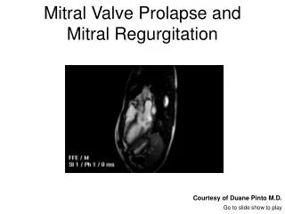
MVP- • Defined as systolic displacement of >2mm of one or both mitral leaflets into the LA below the plane of mitral annulus • Mitral leaflets often thickened >5mm or myxomatous • MVP with thickening of leaflets prone for complications • Prolapse with otherwise normal leaflets and no MR –low risk
Rheumatic MR-commissural fusion,chordal fusion and shortening • Infective endocarditis-leaflet destruction,perforation or deformity • Marfan syn.-long redundant antr.leaflet,aortic pathology • Ischemic MR-restricted leaflet motion • Papillary muscle rupture-a/c MI • Functional MR-annular dilatation • Mitral annular calcification-impair systolic contraction leading to MR
LV dilatation • LA dilatation • Decline in LV contractility • PA pressure from TR jet