Download

1 / 52
590 likes | 933 Vues
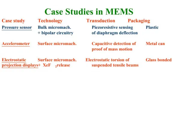
CASE STUDIES IN IMMUNOLOGY. NIRAJ PATEL, MD, MS INFECTIOUS DISEASES AND IMMUNOLOGY. Case 1. 7-year-old girl is brought in by her father for evaluation of full body rash and swelling Symptoms present for 2 weeks

E N D
CASE STUDIES IN IMMUNOLOGY NIRAJ PATEL, MD, MS INFECTIOUS DISEASES AND IMMUNOLOGY
Case 1 • 7-year-old girl is brought in by her father for evaluation of full body rash and swelling • Symptoms present for 2 weeks • After obtaining a careful history and performing a physical examination, you determine that the child has urticaria
Case 1 (cont) • Of the following, the MOST likely cause is A. artificial food coloring B. milk C. new laundry detergent D. shrimp E. upper respiratory tract viral infection
Urticaria vs. Angioedema • Urticaria – superficial dermis • Characterized by intense pruritis due to histamine effect • Angioedema – deeper dermal and subcutaneous layers • May be pruritic but often is a deeper and dull discomfort – burning quality
Acute vs. Chronic Urticaria • Acute Urticaria – lasts 6-8 weeks or less • Viral syndromes (especially in young children) • Insect bites or stings (fire ants, scabies) • Food induced reactions (eat this - get that) • Medication related (antibiotics, NSAIDs, narcotics, angioedema due to ACE inhibitors) • Chronic Urticaria – lasting longer than 8 weeks • Physical urticarias (dermographism, cholinergic, cold) • Urticarial vasculitis • Urticaria/angioedema associated with autoimmunity • Autoimmune urticaria • Idiopathic urticaria
Case 2 • 12 yr old Asian boy presents to clinic with a 3 month history of painful recurring hives • Parents tried keeping a food diary, eliminating Yellow Dye Number 5, changing laundry detergents, bath soaps and shampoos and nothing has helped • Taking cetirizine 10 mg daily which keeps him hive-free. He is otherwise healthy but parents are requesting a referral to identify the cause of the hives.
Case 2 (cont) What is the most likely cause? A. Viral illness B. Allergy to dust mites C. Allergy to soy D. Autoimmune vasculitis
Case 2 answer • Answer is D. The most common cause of chronic (>6 weeks in duration) hives in children is either autoimmune or idiopathic. Signs of a worrisome vasculitis rather than benign urticaria include lesions that are painful instead of itchy; lesions that scar; and lesions that last longer than 72 hours. • Answer choices (A) viral illness, (B) allergy to dust mites, and (C) allergy to soy are most likely to cause acute hives
Acute vs. Chronic Urticaria • Acute Urticaria – lasts 6-8 weeks or less • Viral syndromes (especially in young children) • Insect bites or stings (fire ants, scabies) • Food induced reactions (eat this - get that) • Medication related (antibiotics, NSAIDs, narcotics, angioedema due to ACE inhibitors) • Chronic Urticaria – lasting longer than 8 weeks • Physical urticarias (dermographism, cholinergic, cold) • Urticarial vasculitis • Urticaria/angioedema associated with autoimmunity • Autoimmune urticaria • Idiopathic urticaria
Therapeutic Options • Antihistamines for most with acute short-lasting urticaria • Start with non-sedating, long-acting second generation H1 antagonists (Allegra, Zyrtec, Claritin) and supplement with short-acting, sedating H1 antagonists prn. • Combination therapy with H2 if H1 antagonists do not suffice (30% of cases) • Steroids and other immunosuppressants reserved for severe urticaria associated with angioedema of oropharnyx or other systemic signs, moderate to severe drug reactions, urticarial vasculitis, and refractory cases of CIU
Case 3 • A 17 year old is about to leave for college and she has marked in her chart that she is allergic to penicillin • When you try to question her about this history, she states her mom told her that she had a rash when she was 2 years old and received a course of amoxicillin
Case 3 (cont) Which of the following is the BEST statement regarding penicillin allergy? A. She should continue to avoid all PCN and cephs as there is 80% chance she will have an adverse reaction upon repeat exposure B. She should be referred to an allergist for skin testing to penicillin and possible oral challenge C. Order RAST to PCN to assess likelihood of being persistently allergic to PCN D. Tell her that oral medications are more likely to cause adverse reactions, so only IV, IM forms of PCN she be administered in the future E. She should try to find out more history from her mother
Case 3 answer • Answer is B. Incidence of drug IgE-mediated reaction is ≤10%. Most clinicians would continue avoiding this drug class based on history. Concomitant viral infections or ill-defined immune mechanisms are most common cause of such rashes. Cephs with a side chain unrelated to drug associated with a rash (e.g. amoxicillin) is generally acceptable. 2nd and 3rd generation cephs are less likely to cross react than 1st generation cephs. Overall, cross reactivity rate remains less than 10% for all cephs. • Penicilloyl (major determinant of PCN) is commercially available as skin testing reagent. This, along with Penicillin G is 95% sensitive in detecting persistent PCN allergy. On the other hand, if test is positive, there is a 60% chance of reacting. • Serum sickness, EM, and TEN are non-IgE mediated reactions and cannot therefore be evaluated by immediate hypersensitivity skin testing. If a patient has suffered such a reaction, the general method of management is avoidance.
Cross reacting Antibiotics Modified from Pichichero, Pediatrics 2005; 115:1048-57
Case 4 • 12 yo female presents to ER with 1 week history of an enlarging buttock boil. She has a history of numerous abscesses, including 3 which required incision and drainage. • Hospitalized at 8 yo for Staphylococcus aureus bacteremia and 10 yo for MRSA pneumonia and pneumatocele. Her father also has boils. • Temp is 102, other VSS. PE significant for coarse facial features, a double row of teeth, scoliosis of 20 degree curvature, and numerous scars from prior abscesses. • Laboratories show a normal white blood cell count and normal IgG, IgA, and IgM.
Case 4 The most likely diagnosis is: A. leukocyte adhesion defect B. chronic granulomatous disease C. hyperIgE syndrome D. hyperIgM syndrome E. severe combined immunodeficiency
Case 4 answer • The correct answer choice is (C), hyperIgE syndrome. HyperIgE syndrome, or Job’s syndrome, characterized by recurrent infections usually with Staphylococcus aureus andCandida spp.. Defect is in STAT 3 protein, an intracellular signaling protein important for inflammation. HyperIgE syndrome is autosommal dominant inheritance pattern. Abscesses typically require incision and drainage. HyperIgE is a multisystem disorder. Nonimmunologic characteristics are retained primary teeth, scoliosis, neonatal eczema, broad nasal bridge, coarse facies. The IgE level is typically elevated but can be normal. Treatment is bactrim prophylaxis. • Choice (A), leukocyte adhesion defect (LAD) does result in recurrent staphylococcal infections. However, usually associated with elevated white blood cell count and delayed umbilical cord separation. Associated features of scoliosis and retained primary teeth are not associated with LAD or (B) chronic granulomatous disease (CGD). HyperIgM (D) and severe combined immunodeficiency (E) usually present with hypogammaglobulinemia, particularly IgG and IgA. Severe combined immunodeficiency also usually presents in the first year of life.
http://img.medscape.com/pi/emed/ckb/pediatrics_general/884940-886988-680.jpghttp://img.medscape.com/pi/emed/ckb/pediatrics_general/884940-886988-680.jpg
Case 5 • 15 month old male presents to office with recurrent viral, bacterial infections since birth • Hospitalized twice for severe sepsis and bleeding associated with infections. Worsening peripheral neuropathy for past 3 months • PE normal except for skin which is pale white • Labs: neutropenia and bone marrow smear showing giant inclusion bodies inside WBCs. IgG, IgA, and IgM are normal, and T and B cells are normal in number
Case 5 The most likely diagnosis is: A. Wiskott-Aldrich syndrome B. Chediak-Higashi syndrome C. severe-combined immunodeficiency D. transient hypogammaglobulinemia of infancy E. X-linked agammaglobulinemia
Case 5 answer • The correct choice is (B), Chediak-Higashi syndrome. Chediak-Higashi syndrome is rare and presents in infancy with recurrent viral and bacterial infections. Symptoms include severe infections with Staphylococcus aureus, peripheral neuropathy, bleeding, and oculocuataneous albinism. The defect is in the LYST gene, which assists in lysosomal fusion with the phagosome in WBCs. Defective formation of phagolysosomes results in the inability of white blood cells to lyse bacteria once ingested. This appears as accumulation of “giant inclusion bodies” seen on bone marrow smear, which is the diagnostic method. • Wiskott-Aldrich syndrome (A) features a triad of recurrent sinopulmomary infections, eczema, and thrombocytopenia. Severe combined immunodeficiency (C) usually presents less than 12 months of age with absent IgG, IgA, and IgM and generally a lack of T cells (if B cells are present, they are generally non-functional). Transient hypogammaglobulinemia of infancy (D) is usually associated with a low IgG, and X-linked agammaglobulinemia is associated with panhypogammaglobulinemia (IgG, IgA, IgM).
CHEDIAK-HIGASHI SYNDROME • Autosommal recessive • Impaired phagolysosomal formation • Finding: Giant lysosome granules in phagocytes (neutrophils) • Staphylococcus aureus and viral infections
CHEDIAK-HIGASHI SYNDROME • oculocutaneous albinism • Peripheral neuropathy • Diagnosis: Bone marrow smear • Treatment: bone marrow transplantation, frequent antibiotics
http://img.medscape.com/pi/emed/ckb/dermatology/1048885-1068184-344.jpghttp://img.medscape.com/pi/emed/ckb/dermatology/1048885-1068184-344.jpg
Case 6 • 8 mo male presents to office with history of recurrent pneumonia and otitis media. • Hospitalized at 5 mo of age for pneumonia and bacteremia with Streptococcus pneumoniae. Hospitalized again at 6 mo for Haemophilus influenzae meningitis. He has chronic otorrhea for 2 mo. • PE: well appearing small infant. Weight and length are <5%. Tonsils are absent and there are no palpable lymph nodes.
Case 6 Which of the following is the most likely diagnosis? A. DiGeorge syndrome B. Wiskott-Aldrich syndrome C. X-linked agammaglobulinemia D. hyperIgE syndrome E. chronic granulomatous disease
Case 6 answer • The correct answer is (C) X-linked agammaglobulinemia. X-linked agammaglobulinemia (XLA) occurs in males and characterized by recurrent sinopulmonary infections. Onset of infections occur after 4-6 months of age when maternal immunoglobulin begins to wane. XLA is caused by a mutation in the Bruton’s tyrosine kinase (BTK) gene and results in lack of B cells, cells responsible for producing antibody. Hence, all immunoglobulins IgG, IgA, and IgM are low. Since B cells make up a high proportion of lymphoid tissue including tonsils and lymph nodes, these structures are typically small or absent in patients with XLA. Poor weight gain, along with recurrent infections, should raise suspicion of immunodeficiency. • (A) DiGeorge syndrome associated with features like dysmorphic facies, congenital heart defect, absent thymus, hypocalcemia, and seizures. (B) Wiskott-Aldrich syndrome associated with eczema, thrombocytopenia. (D) HyperIgE (Job’s syndrome) associated with recurrent staphylococcal and candidal infections, and (E) CGD associated with granulomas and recurrent infections with Staphylococcus aureus, Aspergillus fumigatus, and Serratia marscescens most commonly.
Case 7 • A 15 month old male presents to the office with a history of bilateral otorrhea 3 weeks ago. He was hospitalized at 6 and 9 months of age for pneumonia, and bacterial meningitis at 11 months of age. • PE shows a well appearing infant and is otherwise unremarkable. • Laboratory analysis shows IgG <120 and normal IgA and IgM. Antibody to diphtheria, tetanus, and S. pneumoniae are normal. Lymphocyte enumeration reveals normal absolute numbers of T, B, and NK cells.
Case 7 The most appropriate management for this child is: A. intravenous immunoglobulin B. prophylactic antibiotics C. initiate immunization to encapsulated bacteria D. bone marrow transplant E. avoid live viral vaccines
Case 7 answer • Correct answer choice is (B), prophylactic antibiotics. Infant has transient hypogammaglobulinemia of infancy (THI). THI occurs between 1-2 years of age and characterized by recurrent bacterial infections. Most likely etiology is prolonged period for physiologic B-cell function. THI is transient due to presence of B cells. Distinguished from XLA where no B cells are present. In XLA, IgG, IgA, and IgM are all low. In THI, IgG is low but IgA, IgM are normal. Treatment for THI is prophylactic antibiotics (B). Patients with THI usually reach normal antibody levels by 2-4 years of age. • IVIG (A) is treatment of choice for XLA but can inhibit antibody production in THI and is not usually indicated as treatment. Patient has normal antibody responses to childhood vaccines, and additional immunizations to encapsulated bacteria (C) would not be warranted. Bone marrow transplantation (D) is treatment of choice for severe combined immunodeficiency and other T cell disorders, not THI. Patients who have THI are recommended to receive their full childhood immunization series (E), although antibody response to vaccine may be diminished.
Antibody Deficiency Syndromes *Adapted from New Orleans Pediatric Board Review, Ricardo Sorensen
Case 8 • A 3 year old female presents to the office for a well child check up. • She began to lose motor milestones at age 2 years, and has a history of repeated sinusitis and pneumonias often requiring hospitalization and intravenous antibiotics. Recently, the child has had difficulty maintaining gait while walking and the parents report the child as stumbling frequently. • A paternal grandmother died at age 24 from severe pneumonia. • Physical examination is remarkable for several prominent vessels under the skin and eye.
Case 8 The most likely diagnosis is: A. leukocyte adhesion deficiency B. DiGeorge syndrome C. common variable immunodeficiency D. chronic granulomatous disease E. ataxia-telangiectasia
Case 8 answer • The correct answer choice is (E), ataxia-telangiectasia. Ataxia-telangiectasia (AT) is an autosommal recessive disorder. Although ataxia and telangiectasias are unique features associated with this disorder, the initial manifestation of AT is lost of motor milestones. AT is associated with defective DNA damage repair. Therefore, patients with AT should avoid radiation (such as certain imaging modalities like CT) due to DNA breakage which cannot be repaired. An elevated alpha fetal protein suggests the diagnosis of AT. Treatment is by bone marrow transplant. • The features in this child are not associated with the remaining answer choices (A-D).
http://www.google.com/imgres?imgurl=http://geneticpeople.com/wp-content/uploads/2010/02/Ataxia-telangiectasia1.gif&imgrefurl=http://geneticpeople.com/%3Ftag%3Dmutation%26paged%3D4&usg=__p8KxhlmyOhBtsJFtpiisbsQexps=&h=526&w=652&sz=276&hl=en&start=1&zoom=1&um=1&itbs=1&tbnid=wIgIKiXGxTMpVM:&tbnh=111&tbnw=138&prev=/images%3Fq%3Dataxia%2Btelangiectasia%26um%3D1%26hl%3Den%26safe%3Dactive%26tbs%3Disch:1http://www.google.com/imgres?imgurl=http://geneticpeople.com/wp-content/uploads/2010/02/Ataxia-telangiectasia1.gif&imgrefurl=http://geneticpeople.com/%3Ftag%3Dmutation%26paged%3D4&usg=__p8KxhlmyOhBtsJFtpiisbsQexps=&h=526&w=652&sz=276&hl=en&start=1&zoom=1&um=1&itbs=1&tbnid=wIgIKiXGxTMpVM:&tbnh=111&tbnw=138&prev=/images%3Fq%3Dataxia%2Btelangiectasia%26um%3D1%26hl%3Den%26safe%3Dactive%26tbs%3Disch:1
http://images.google.com/imgres?imgurl=http://www.nature.com/embor/journal/v5/n8/images/7400210-f1.jpg&imgrefurl=http://www.nature.com/embor/journal/v5/n8/fig_tab/7400210_f1.html&usg=__8gzOaxOJqqZgNeph_5F8VYDJLd8=&h=597&w=500&sz=28&hl=en&start=1&um=1&itbs=1&tbnid=Ja_2I7dRGhl_xM:&tbnh=135&tbnw=113&prev=/images%3Fq%3Dataxia%2Btelangiectasia%26um%3D1%26hl%3Den%26safe%3Dactive%26tbs%3Disch:1http://images.google.com/imgres?imgurl=http://www.nature.com/embor/journal/v5/n8/images/7400210-f1.jpg&imgrefurl=http://www.nature.com/embor/journal/v5/n8/fig_tab/7400210_f1.html&usg=__8gzOaxOJqqZgNeph_5F8VYDJLd8=&h=597&w=500&sz=28&hl=en&start=1&um=1&itbs=1&tbnid=Ja_2I7dRGhl_xM:&tbnh=135&tbnw=113&prev=/images%3Fq%3Dataxia%2Btelangiectasia%26um%3D1%26hl%3Den%26safe%3Dactive%26tbs%3Disch:1
Case 9 • A 9 year old female presents to the office with recurrent sinusitis since age 4. • She has had 3 pneumonias in the same time period which were successfully treated with antibiotics. • PE is unremarkable. Laboratory analysis shows IgG 800, IgM 150, and IgA <6. Antibody to diphtheria, tetanus, and S. Pneumoniae are within the normal range. Lymphocyte enumeration shows normal T, B, and NK cells.
Case 9 Which of the following should most likely be avoided? A. live viral vaccines B. obtaining sinus culture results C. allergy testing to environmental (indoor and outdoor) allergens D. intravenous immunoglobulin E. prophylactic antibiotics
Case 9 answer • Correct choice is (D), intravenous immunoglobulin. Patient has selective IgA deficiency, most common primary immunodeficiency (frequency ~ 1:400-1:500 persons). Selective IgA deficiency presents with recurrent sinopulmonary infections. IgG, IgM are normal, and IgA absent. IVIG (D) contains small amounts of IgA. Some patients with selective IgA deficiency have preformed antibodies to IgA resulting in anaphylactic reaction when given IVIG. • Patients with selective IgA deficiency tolerate live viral vaccines (A). Sinus culture results (B) would direct antibiotic therapy in the future, and allergy testing (C) may reveal other factors for increased risk for sinusitis. Prophylactic antibiotics (E) may be helpful in patients with recurrent sinusitis or bronchitis.
Case 10 • 6 month old Caucasian male infant presents to the Emergency Room with a 6 day history of pallor, fussiness, hypoxia, and pulmonary infiltrates. • Past medical and family history are non-contributory. • On physical exam, the infant is ill-appearing, tachypneic and is less than the 10% for weight. Laboratory analysis shows an IgG of 16, IgA <6, and IgM of 350. Antibody to diphtheria, tetanus, and S. Pneumoniae are absent. Lymphocyte enumeration shows normal number of T, B, and NK cells and normal function. HIV PCR is negative.
Case 10 The most likely diagnosis is: A. severe combined immunodeficiency B. X-linked agammaglobulinemia C. HIV infection D. DiGeorge syndrome E. hyperIgM syndrome