Download
1 / 7
70 likes | 220 Vues
Quantitative Comparison of Conventional and Oblique MRI for Detection of Herniated Discs. Automatic Herniation Detection. A collaborative project with Doug Dean Erin Hannen. Purpose. Development of an algorithm for: S egmentation of individual spinal disks

E N D
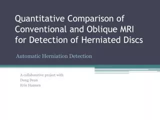
Quantitative Comparison of Conventional and Oblique MRI for Detection of Herniated Discs Automatic Herniation Detection A collaborative project with Doug Dean Erin Hannen
Purpose • Development of an algorithm for: • Segmentation of individual spinal disks • Determination of specific, quantitative properties of each disc • Use properties to determine if a disc is herniated or normal
Approach • Modify methods from “Desiccation diagnosis in lumbar discs from clinical MRI with a probabilistic model” • Intensity: Obtain histogram. Herniated discs typically have lower intensity profile due to spreading of the nucleus pulposus over a larger area. Individual intensity values and the average intensity value are obtained
Approach 2. Probabilistic Model: Z[n] = normalization factor β1 ,β2= tuning parameters UA = appearance parameter, Determined by intensity values, averaged intensity, and a defined pixel neighborhood US =shape parameters Determined based on location coordinates from points that define shape of the disk
Current Progress • Lumbar discs segmented • Semi-automated • Edge detection, MATLAB image processing tools • Disc location defined • Centroid of segmentation boundary calculated • Overlay of segmentation boundary onto original image • Average intensity over entire segmented disk
I = 80.400 I = 86.4614 I = 84.6678 I = 70.1894
Project timeline • April 12: First project presentation • April 13-27: Continue reading literature articles comparing methods for disc quantification. Begin writing MATLAB code for herniation detection using data from class labs or phantom images. • April 28: Mid project presentation • April 28-May 15: • Refine segmentation methods, where needed • Finish developing herniation detection code • Ensure successful implementation using acquired MRI data • May 16 & 17: Final project presentation