Acute Kidney Injury and Hyperkalaemia
500 likes | 1.41k Vues
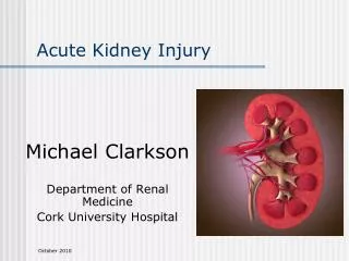
Acute Kidney Injury and Hyperkalaemia. Ben Ryan Final Year Medical Student. Contents. Fluid and Electrolyte Requirements A little bit on prescribing fluids Hydration Status Exam U rine output Acute Kidney Injury Definition and Diagnosis Causes Approach to Management

Acute Kidney Injury and Hyperkalaemia
E N D
Presentation Transcript
Acute Kidney Injury and Hyperkalaemia Ben Ryan Final Year Medical Student
Contents • Fluid and Electrolyte Requirements • A little bit on prescribing fluids • Hydration Status Exam • Urine output • Acute Kidney Injury • Definition and Diagnosis • Causes • Approach to Management • Drugs to stop in AKI • Hyperkalaemia • Definition • Causes • Management • Uraemia • ABG interpretation
Why is this important? • You will see Acute Kidney Injuries very commonly in hospitals • Important to have a good grasp on it’s clinical significance • AKI can significantly change medication • Fluid prescribing is an extremely common job for FY1s • Very easy for questions on this topic to come up on progress
Fluid and Electrolyte Requirements • The average person requires the following per day: • ~25mL/kg/day of water per day. • So if they were 70kg, 1750mL per day • Slightly less needed if elderly/frail/cardiac impaired/malnourished • Potassium, sodium and chloride: • 1mmol/kg/day • If they were 70kg, that would be 70mmol/kg/day • 50-100g of glucose per day
Hydration Status • Hydration Status is a genuine examination for Year 3 OSCE • Too much fluids? • May lead to pulmonary oedema (fluid on the lungs) and general swelling • Worse in people with a history of cardiac failure and renal disease • Too much fluids can also cause hyponatraemia (many things can cause hyponatraemia however!) • Too little fluids: • May feel unwell, low urine output, low kidney function, hypovolaemic shock
Hydration Status Exam • As always: • General inspection – IV fluids? Catheter? Fluid balance charts? Patient look well? Look dehydrated? Short of breath? • Hands, arms - capillary refill and skin turgor • Head and neck • Sunken eyes – dehydration • Dry mouth • JVP – increased in fluid overload • Chest – listen to heart and lungs – most important to listen for crackles at base of lungs (pulmonary oedema)
continued • Abdomen • Ascites? • Legs • Peripheral oedema • Offer full history, U&Es, observations and fluid balance chart • Can offer serial weights to look for quick change in weight (often due to change in fluid levels) • Most important to think about signs of dehydration or fluid overload, clinical items like fluid balance charts and catheters, and do a quick review of chest and abdo
Prescribing Fluids • Think about what their daily requirements are based on weight • Think about whether they are dehydrated or fluid overloaded • Are they eating and drinking enough? At all? • Do they have cardiac failure of renal failure? • Fluids are prescribed in their own section on a prescription chart • Why do they need fluids? Quick resuscitation? Daily maintenance fluids to prevent dehyrdration?
Prescribing Fluids • Some different fluids: • 0.9% sodium chloride solution – commonly prescribed • Hartmann’s – supposed to be more like plasma • 5% glucose • Also potassium is often added to fluids before being given to ensure potassium requirements met
Prescribing Fluids • Maintenance fluids (let’s pretend they are Nil By Mouth and need to meet their daily requirements) • Most patients would get ‘2 sweet, 1 salty’ in a day • This means that over a 24 hour period they will be prescribed 3 bags of fluids • 2 bags will be 5% glucose • The other bag will be salty – 0.9% sodium chloride or Hartmann’s • For example: 1 Litre of 5% glucose, 500mL of 0.9% sodium chloride, 500mL or 5% glucose • You could add potassium additives to these bags to make sure they get their daily requirements of potassium • When prescribing fluids, you decide how long it should take for the bag to be given to them (the rate)
Summary • ~25ml/kg/day of water – in a 70kg this is 1750mL • 1mmol/kg/day of potassium, sodium and chloride • If maintenance fluids required, normally 2 sweet bags and 1 salty • I’ve seen a patient be very unwell from fluid overload because of inappropriate fluid prescribing – always check fluid balance charts and make sure they are getting what’s appropriate
Urine Output • Urine output is monitored on fluid balance charts • Best recorded by use of a catheter – other ways too however like urine bottles • If urine output is less than 0.5mL/kg/hour = oliguria and a sign of acute kidney injury • A 70kg person should be making at least 35mL of urine per hour, 840mL per day
Acute Kidney Injury (AKI) • A decline in renal excretion function that can result in failure to maintain fluid,electrolyte and acid-base homeostasis • Causes can be pre-renal, renal or post-renal • Diagnosed by a rise in blood creatinine levels or low urine output • Staged from 1-3 based on blood creatinine/urine output • AKI has important clinical consequences • Changes in medications, affects prognosis, and will likely mean admission in hospital • Can cause hyperkalaemia, pulmonary oedema, metabolic acidosis and uraemia
Causes – Pre-renal • Hypovoleamia (reduces kidney excretory function because less blood flow to kidneys) • Lots of things can cause hypovolaemia • Reduced cardiac output • Cardiac failure, sepsis, drugs
Causes - Renal • Drugs • ACE inhibitors, NSAIDs • Vascular causes • Vasculitis, thrombosis • Glomerular damage • Glomerulonephritis • Tubular damage • Can be secondary from rhabdomyolysis, myeloma and contrast • Interstitial • Interstitial nephritis • I haven’t got a good grasp on many of these causes so don’t worry too much about them – you’ll do nephrotic syndrome and nephritic syndrome which will cover some of these - most important thing to be aware of is drugs that affect the kidneys
Causes – post-renal • Obstruction: • Renal stones • Blocked catheters • Enlarged prostate • Literally anything that will block the urinary tract
Diagnosis • Everybody who comes into hospital will have U&Es done, which has creatinine levels on it • Creatinine is excreted in urine and isn’t absorbed anywhere in the nephron so it’s a good marker of the kidney’s ability to form ultrafiltrate • Looking at creatinine levels (compared to their previous creatinine levels) is the main way AKI is diagnosed • Important to look at their previous/most recent U&Es so it can be compared • You can also use urine output
Diagnostic criteria • Rise in serum creatinine of >26 micromol/L within 48 hours • Or a >50% rise in serum creatinine within the past 7 days • Or a fall in urine output to less than 0.5ml/kg/hour for more than 6 hours • In reality however, this is what you should be looking for: • Creatinine significantly higher than it was previously • Low urine output • Lower eGFR – estimated eGFR, uses creatinine to work it out anyhow (although they don’t appear to be using this in the actual diagnostic criteria of AKI as much? Might change in the future)
Staging – not going in go into specifics because it’s too much • NICE has 3 stages:stage 1 – mild increase in creatinine or low urine output for 6 hoursstage 2 – moderate increase in creatinine or low urine output for 12 hoursstage 3 – severe increase in creatinine of low urine output for 24 hours • There is other criteria like RIFLE • Mainly just looks at changes in creatinine, urine output and eGFR. Identified as Risk, Injury, Failure, Loss of function, or End stage renal disease (RIFLE)
Approach to Management • Identify cause: • Pre-renal – try to identify possible causes of reduced blood flow to the kidneys. Loss of fluids like vomiting? Sepsis? Cardiac failure? • Renal – changes to medications? Protein in urine? Blood in urine? Raised Urinary Sodium? • Post-renal – urinary tract symptoms, nitrites in urine? • Treat underlying cause: • Pre-renal – might mean giving fluids or treating cause of poor blood flow to kidneys • Renal – stopping appropriate medications (regardless of cause), treating cause of renal damage • Post-renal – relieving obstruction
Approach to Management of AKI • Often urgent same day admission if not already in hospital • If high risk, discuss with renal team • Need to be very careful with medication and fluid prescriptions • Many medications can make AKI worse, or the low renal function can cause increased toxicity of drug
NSAIDs:Ibuprofen, naproxen, diclofenac Aminoglycosides:Gentamicin ACE inhibitors:an interesting one in kidneys, they are helpful in hypertension + CKD, but they are bad for kidneys in AKI Diuretics:Furosemide and others Drugs and AKI
Quick Summary • Potassium levels above 6mmol/L is an emergency • Can predispose to cardiac arrhythmias and cardiac arrest • Can be caused by medications, poor kidney function, endocrine abnormalities, and release of potassium from muscles or tumours • Requires an immediate ECG (can cause ECG changes) • Requires immediate treatment which should be memorised • IV calcium gluconate to stabilise cardiac membrane • Salbutamol nebulisers, insulin and glucose to reduce potassium levels
Potassium Levels • Normal is 3.5-5 mmol/L • Mild hyperkalaemia is 5-6 mmol/L • Not an emergency but would probably want an ECG still • Emergency if potassium above 6mmol/L • Potassium levels may just appear raised because of faulty blood test • ‘Pseudohyperkalaemia’ • Haemolysis of blood during venepuncture • Excessive tourniquet or fist clenching
Potassium and the Heart • Can cause arrhythmias and cardiac arrest • Can cause ECG changes indicative of hyperkalaemia: • (least severe) Prolonged PR interval • Tall tented T waves • Widening of QRS complex • (most severe) Can appear to look like a sin wave (y = sin x)
Causes • Decreased kidney function • Medications (spironolactone in particular) • Aldosterone deficiency (Addison’s disease = adrenal insufficiency, aldosterone is one of the adrenal hormones) • Potassium release from cells (rhabdomyolysis, burns, tumour lysis syndrome
Management • Above 6: • ECG and repeat potassium • If ECG changes or potassium above 6.5: • Contact renal registrar on call • Monitor cardiac rhythm – ECG monitor, different to a 12-lead ECG • Drug 1: IV calcium gluconate • 10% 10mL given over 5 minutes. Can repeat every 5 minutes to a maximum of 3 doses • Drug 2: 10 units of Short-acting insulin (Actrapid) and 50% Glucose 50mL • Drug 3: Salbutamol 10mg Nebulised • ?Drug 4?: Consider Sodium Bicarbonate if acidotic
Uraemia • Azotemia – high levels of blood urea • Uraemia – azotemia + pathological features • Uraemia can affect many systems. Some features to look out for:reduced CNS functionItchingHiccupsgenerally unwell • Caused by AKI and poor kidney function
ABG Interpretation • Normal ranges:pH – 7.35-7.45pO2 – 10-14pCO2 – 4.5-6 (high co2 causes acidosis)Base excess: -2 – 2HCO3: 22-26 (high HCO3 causes alkalosis) • Will also show electrolytes and lactate
ABG definitions • Acidosis – low pHAlkalosis – high pH • Metabolic – renal/GI - referring to HCO3 and base excess usually • Respiratory – referring to CO2 and oxygen • Compensation – one system has altered it’s levels to counter the change in pH – if there is a respiratory acidosis from high CO2, kidneys may retain more HCO3 to compensate (respiratory acidosis with metabolic compensation) • Respiratory failure = low oxygen type 1 – CO2 is normal/low type 2 – CO2 is high
Approach • Check pH first – normal, acidotic or alkalotic? • If altered, establish why. Would the CO2 explain it? Would the HCO3? • Is the other system compensating for it? Is it fully compensating for it, or just partially? • Check oxygen – normal, or low? • Low = respiratory failure. Check CO2.CO2 raised means type 2 respiratory failure. Otherwise is type 1 respiratory failure.
There is also this thing called the ‘Anion gap’ which is used to assess metabolic acidosis • I have never heard anybody mention it in clinical practice, progress test or OSCEs • I don’t especially like it and people seem to calculate it differently • I’ll mention it briefly but no need to stress about it
Anion Gap in Metabolic Acidosis • If there is a metabolic acidosis, the anion gap will help us in our evaluation • Raised anion gap means acid is being produced or ingested • Normal anion gap means loss of HCO3Anion gap = cations (sodium, potassium) minus anions (chloride, bicarbonate). Normal range depends on how it was calculated • Raised anion gap: (acids from sources such as ketones or lactate) • DKA, lactic acidosis, sepsis • Normal anion gap: (loss of HCO3 often to do with kidneys or GI)
MCQ 1 • A 67 year old male presents to the emergency department with palpitations. A blood test shows that his potassium is raised at 6.7. Which of the following would least appropriate as part of your management plan? • A – IV Calcium Gluconate • B – 12-lead ECG • C – ECG monitor • D – Insulin + Glucose • E – Salbutamol nebulizers • F – Repeat the blood test and ensure patient fist is clenched
MCQ 2 • A 67 year old male presents to the emergency department with palpitations. A blood test shows that his potassium is raised at 6.7. An ECG has been performed. Which of the following would you least expect to see? • A – Depressed T waves • B – Broad QRS complex • C – Sinusoidal pattern • D – Prolonged PR interval • E – Arrhythmias
MCQ 3 • A 67 year old male presents to the emergency department with palpitations. A blood test shows that his potassium is raised at 6.7. An ECG shows tall tented T waves and a broad QRS. Which of the following would be your next step in management? • A – IV Calcium Gluconate • B – Spironolactone • C – Sodium Bicarbonate • D – Insulin + Glucose • E – Salbutamol nebulizers
MCQ 4 • A 67 year old female with a history of iron-deficiency anaemia and Burkitt’s lymphoma, for which she is receiving chemotherapy, presents to the emergency department with palpitations. A blood test shows a raised potassium at 6.7 and creatinine is markedly raised. ECG shows tall tented T waves. She has recently finished a cycle of chemotherapy. Her observations are normal. Which of the following is the most likely cause of her blood test findings? • A – Spironolactone usage • B – Addison’s Disease • C – Hypovolaemic shock • D – Tumour Lysis Syndrome • E – Rhabdomyolysis
MCQ 5 • A 58 year old female is found to have markedly raised creatinine compared to baseline. She recently been started on an anti-hypertensive medication. Which of the following medications would best explain this finding? • A – Ramipril • B – Ibuprofen • C – Paracetamol • D – Amlodipine • E - Bisoprolol
MCQ 6 • A 72 year old male presents to the emergency department feeling generally unwell with fevers and a productive cough. His blood pressure is 88/56, his respiratory rate if 24/minute, and he is slightly confused. His urine dipstick was negative for leukocytes, nitrites and blood. He did not complain of any urinary symptoms. His serum creatinine is markedly raised. What is the most likely cause of his AKI? • A – Cardiac failure • B – Septic Shock • C – Kidney Stones • D – Benign Prostatic Hyperplasia • E – Nephritic Syndrome
MCQ 7 • A 72 year old female is an in-patient on a surgical ward. She is known to have chronic kidney disease and a low BMI. She is currently suffering from severe diarrhoea and vomiting. She takes Ramipril and spironolactone for heart failure. She is currently having palpitations and has called for help. You are part of the team that has been called. You review her fluid prescription chart for the past 24 hours:500mL of 5% glucose with 20mmol of potassium over 6 hours500mL of 5% glucose with 20mmol of potassium over 6 hours1 litre of Hartmann’s with 40mmol of potassium additive over 12 hoursWhich of the following is the most likely cause of her palpitations? • A – DehydrationB – HyperkalaemiaC – Fluid overloadD – SespisE – Hyponatraemia
MCQ 8 • A 72 year old female is a 50kg in-patient on a medical ward after a recent myocardial infarction. She is known to have end-stage kidney disease. She has became progressively short of breath over the past few hours and has called for help. She has no pain anywhere, but has complained of bilateral leg swelling. The nurse tells you that she was feeling well previously, and was sat up in her bed eating and drinking. Upon arriving at the scene you review her fluid prescription chart for the past 24 hours:500mL of 5% glucose with 20mmol of potassium over 6 hours500mL of 5% glucose with 20mmol of potassium over 6 hours1 litre of 0.9% sodium chloride over 12 hoursWhich of the following do you feel is the most likely cause of her shortness of breath? • A – Hypokalaemia • B – Pulmonary Oedema • C – Uraemia • D – Sepsis • E – Pulmonary Embolus
MCQ 9 • 25 year old with severe nausea and vomiting for 24 hours. ABG shows: • pH – 7.55 (7.35-7.45)pO2 – 12 (10-14)pCO2 – 5.2 (4.5-6)Base excess: 5 (-2 – 2)HCO3: 30 (22-26) • Describe
MCQ 10 • 58 year old male with COPD presents to clinic. ABG results show: • pH – 7.33 (7.35-7.45)pO2 – 8 (10-14)pCO2 – 7 (4.5-6)Base excess: 5 (-2 – 2)HCO3: 30 (22-26) • Describe
MCQ 11 • 19 year old male presents with acute shortness of breath. ABG results show: • pH – 7.43 (7.35-7.45)pO2 – 8 (10-14)pCO2 – 4.6 (4.5-6)Base excess: 0 (-2 – 2)HCO3: 24 (22-26) • Describe
MCQ 12 • 7 year old male presents with lethargy, weight loss and urinary frequency. Is currently feeling very unwell. ABG results show: • pH – 7.25 (7.35-7.45)pO2 – 11 (10-14)pCO2 – 5 (4.5-6)Base excess: -6 (-2 – 2)HCO3: 18 (22-26) • Describe