Exercise Stress Electrocardiography
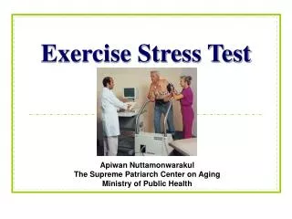
Exercise Stress Electrocardiography. Dr Bijilesh.U. Exercise is a common physiological stress used to elicit cardiovascular abnormalities not present at rest and to determine adequacy of cardiac function.

Exercise Stress Electrocardiography
E N D
Presentation Transcript
Exercise Stress Electrocardiography Dr Bijilesh.U
Exercise is a common physiological stress used to elicit cardiovascular abnormalities not present at rest and to determine adequacy of cardiac function. Exercise ecg - one of the most frequent noninvasive modalities used to assess patients with suspected or proven cardiovascular disease. Estimate likelihood & extent of CAD , the prognosis , determine functional capacity & effects of therapy.
Exercise physiology Exercise protocols Electrocardiographic measurements Nonelectrocardiographic observations Exercise test indications Specific Clinical Applications Safety and risks of exercise testing Termination of exercise
EXERCISE PHYSIOLOGY • Exercise - body's most common physiologic stress - places major demands on CVS • Exercise considered most practical test of cardiac perfusion and function • Fundamentally involves the measurement of work • Common biologic measure of total body work is oxygen uptake • Cardiac output can increase as much as six-fold
EXERCISE PHYSIOLOGY • Acceleration of HR by vagal withdrawal • Increase in alveolar ventilation • Increased venous return- sympathetic venoconstriction. • Early phases - cardiac output increased by augmentation in stroke volume and heart rate • Later phases by sympathetic-mediated increase in HR
During strenuous exertion, sympathetic discharge is maximal and parasympathetic stimulation is withdrawn • Vasoconstriction of most circulatory body systems - except in exercising muscle , cerebral and coronary circulations • Catecholamine release enhances ventricular contractility
As exercise progresses skeletal muscle blood flow is increased O2 extraction increases by as much as threefold total calculated peripheral resistance decreases systolic blood pressure, mean arterial pressure, and pulse pressure increase • Diastolic blood pressure does not change significantly.
V O2 • Total body or ventilatory O2 uptake - amount of O2 extracted from air as the body performs work • Determinants of VO2 - cardiac output - peripheral AV oxygen difference • Maximal AV difference is constant 15 to 17 mL/dL • Vo2 - estimate of maximal cardiac output.
V O2can be estimated fromtreadmillspeedandgrade • Vo2 = (MPH ˣ 2.68 ) ˣ [.1 + ( Grade ˣ 1.8) ] + 3.5 • Vo2 can be converted to METS by dividing by 3.5.
M O2 • Myocardial oxygen uptake is the amount of oxygen consumed by the heart muscle • Determinants of M O2 – Intramyocardial wall tension - Contractility & HR • Mo2 - estimated by - HR & SBP (double product). • Exercise-induced angina often occurs at the same Mo2 • Higher double product - better myocardial perfusion
Maximum heart rate • Maximum heart rate (MHR) : 220 – age • Overestimate maximum heart rate in females MHR = 206 − 0.88 (age in years) • MHR decreased in older persons • Age-predicted maximum heart rate is a useful measurement for safety reasons
Post exercise phase - hemodynamics return to baseline within minutes • Vagal reactivation - important cardiac deceleration mechanism after exercise • Accelerated in athletes but blunted in chronic heart failure
Metabolic Equivalent • Refers to a unit of oxygen uptake in a sitting, resting person • Common biologic measure of total body work is the oxygen uptake • One MET is equated with the resting metabolic rate (3.5 mL of O2/kg/min) • MET value achieved from an exercise test is a multiple of the resting metabolic rate
METS associated with activity = Measured Vo2 / 3.5 (both in mL O2/kg/min) • Measured directly (as oxygen uptake) or estimated from the maximal workload achieved - using standardized equations
Calculationof METs on the Treadmill METs = Speed x [0.1 + (Grade x 1.8)] + 3.5 3.5 Calculated automatically by Device!
Clinically Significant Metabolic Equivalents for Maximum Exercise
Exercise Test Modalities • Isometric, dynamic, and a combination of the two. • Isometric exercise - constant muscular contraction without movement • Moderate increase in cardiac output and only a small increase in vo2 - insufficient to generate an ischemic response. • Dynamic exercise - rhythmic muscular activity resulting in movement
Exercise Protocols • Dynamic protocols are most frequently used to assess cardiovascular reserve • Should include a low-intensity warm-up and a recovery or cool-down period • Optimal for diagnostic and prognostic purposes - Approximately 8 to 12 minutes of continuous progressive exercise - myocardial oxygen demand elevated to patient's maximum
Arm Ergometry • Bicycle Ergometry • Treadmill Protocol • Walk Test
Arm Ergometry • Involve arm cranking at incremental workloads of 10 to 20 watts for 2- or 3-minute stages HR & BP responses to a given workload > leg exercise Peak vo2 and peak HR - 70% of leg testing Bicycle Ergometry • Involve incremental workloads starting at 25 – 50 watts • Lower maximal VO2 than the treadmill
Treadmill Protocol s • Bruce • Modified Bruce • Naughton and Weber • ACIP (Asymptomatic cardiac ischemia pilot trial) • Modified ACIP
Tread mill protocolBruce multistage maximal treadmill protocol • 3 minutes periods to achieve steady state before workload is increased • Limitation - relatively large increase in vo2 between stages • Modified Bruce protocol - Older individuals or those whose exercise capacity is limited • Modified by two 3 min warm up stages at 1.7mph % 0 % grade and 1.7mph % 5%grade.
Naughton and Weber protocols use 1-2min stages with 1-MET increments between stages • Asymptomatic cardiac ischemia pilot trial and modified ACIP protocols use 2min stages with 1.5mets increments between stages - after two 1min warm up • Functional capacity overestimated by 20% -if handrail support is permitted
Walk Test • A 6-minute walk test or a long-distance corridor walk • Provide an estimate of functional capacity in patients who cannot perform bicycle or treadmill exercise • Older patients ,heart failure, claudication, or orthopedic limitations • Walk down a 100-foot corridor at their own pace - cover as much ground as possible in 6 minutes • Total distance walked is determined and the symptoms experienced by the patient are recorded.
Cardiopulmonary Exercise Testing • Involves measurements of respiratory oxygen uptake (vo2) , carbon dioxide production ( vco2 ) and ventilatory parameters during a symptom- limited exercise test • Patient wears a nose clip and breathes through a nonrebreathing valve
Technique No caffeinated beverages or smoke 3hr before Wear comfortable shoes and clothes. Unusual physical exertion should be avoided Brief history & physical examination performed Explain risks and benefits Informed consent is taken
12 lead ECG is recorded with electrodes at the distal extremities Torso ECG is obtained in supine & standing position If false +ve test is suspected, hyperventilation should be performed
Room temp should be 18 –24 C & humidity < 60% Walking should be demonstrated to the patient HR, BP & ECG recorded at end of each stage. Resuscitator cart, defibrillator and appropriate cardioactive drugs should be available
Optimal patient position in the recovery phase ? supine • Sitting position, less space is required and patients are more comfortable • Supine position increases end-diastolic volume and has the potential to augment ST-segment changes
Lead system Mason-Likar modification • Modification of the standard 12-lead ECG • Extremity electrodes moved to torso to reduce motion artefact • Results in right axis shift increased voltage in inferior leads loss of inferior Q waves new Q waves in lead aVL
Types of ST-Segment Displacement • J point, or junctional, depression - normal finding in exercise • In myocardial ischemia, ST segment becomes horizontal, • With progressive exercise depth of ST segment may increase • In immediate post recovery phase ST segment displacement may persist with down sloping ST segments and T wave inversion - returning to baseline after 5-10 min • In 10% , ischemic response may appear in recovery phase
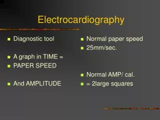
Measurement of ST-Segment Displacement PQ junction is chosen as isoelectric point TP segment is true isoelectric point but impractical choice Abnormal ST depression 0.1mv (1mm) or > ST depression from PQ junction with a flat ST segment slope ( <0.7-1mv /sec) 80 msec after J point (ST 80) in 3 consecutive beats with a stable base line
When ST 80 measurement difficult at rapid heart rates > 130/mt measure at ST 60 When ST is depressed at rest- additional 0.1mv or more during exercise is considered abnormal
1.PQ JUNCTION 2. J POINT 3.ST 80
Upsloping ST segment Rapid upsloping ST segment (more than 1 mV/sec) depressed less than 1.5 mm after the J point - normal
Slow upsloping ST segment at peak exercise In patients with high CAD prevalence, slow up sloping ST ,depressed > 1.5mm ST 80 is considered abnormal
ST segment elevation • 0.1mv ( 1mm) or greater of ST elevation, at ST 60 in 3 consecutive beats - abnormal response. • More frequently with AWMI - early after event - decreases in frequency by 6 weeks • ST elevation is relatively specific for territory of ischemia
In leads with abnormal Q waves - not a marker of more extensive CAD and rarely indicates ischemia. • When it occurs in non q wave lead in a patient without previous MI - transmural ischemia • In a patient who has regenerated embryonic R waves after AMI - significance similar
Eight typical exercise ecg patterns at rest and at peak exertion
T Wave Changes • Transient conversion of a negative T wave at rest to positive T wave in exercise – pseudonormalisation • Nonspecific finding in patients without prior MI • Does not enhance diagnostic or prognostic content of test
Nonelectrocardiographic Observations • Blood pressure • Maximal Work Capacity • Heart rate response • Heart Rate Recovery • Chest discomfort • Rate-Pressure Product
Blood pressure • Normal exercise response - increase SBP progressively with increasing workloads. • Range from 160 to 200 - higher range in older patients with less compliant vessels • Abnormal • Failure to increase SBP > 120 mm Hg • Sustained decrease greater than 10 mm Hg • Fall in SBP below resting values • Diastolic BP doesn’t change significantly
Conditions other than myocardial ischemia associated with abnormal BP response Cardiomyopathy Cardiac arrhythmias LVOT obstruction Antihypertensive drugs Hypovolemia • An exaggerated BP increase with exercise - increased risk of future hypertension
Maximal Work Capacity • Important prognostic measurement of exercise test • Limited exercise capacity - increased risk of fatal and nonfatal cardiovascular events • In one series - adjusted risk of death reduced by 13% for each 1-MET increase in exercise capacity • Estimates of peak functional capacity for age and gender - known for most protocols
Heart rate response • Sinus rate increases progressively with exercise. • Inappropriate increase in heart rate at low work loads - • Atrial fibrillation • Physically deconditioned • Hypovolumic • Anemia • Marginal left ventricular function
Chronotropic incompetence • Decreased heart rate sensitivity to the normal increase in sympathetic tone during exercise • Inability to increase heart rate to at least 85%of age predicted maximum. • Associated with adverse prognosis
Heart Rate Recovery(HRR) • Abnormal HRR refers to a relatively slow deceleration of heart rate following exercise cessation • Reflects decreased vagal tone - associated with increased mortality • Value of 12 beats/min or less - abnormal