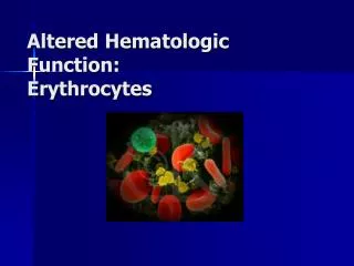
Hematologic examination
Hematologic examination. Jan Živný , Martin Vokurka , Stanislav Matou šek Department of Pathophysiology. Clinical signs of hemato logy diseases. Anemi a → hypoxi a signs – tiredness, weakness, lack of breath, paleness → c ardiovas c ul ar s ymptoms – palpitation

Hematologic examination
E N D
Presentation Transcript
Hematologic examination Jan Živný, Martin Vokurka, Stanislav Matoušek Department of Pathophysiology
Clinical signs of hematology diseases • Anemia → hypoxia signs – tiredness, weakness, lack of breath, paleness • → cardiovascularsymptoms – palpitation • Polycythemia→ hyperviscoseblood → risk of thrombosis • Bleeding, spontaneous bleeding, continuous bleeding • Thrombosis → embolia – symptoms depend on the localisation – DVT, pulmonary embolism • Frequent infections
Hematologic disease • Leukemias etc. • Myelo-(proliferative) • Lympho- • (granulo-, mono-) • Disorders of Red Cells • Anemias • Hemostatic disorders • Primary hemostasis • Coagulation
MAIN REASONS OF HYPOXIA • Lack of oxygen in inspired air / low oxygen partial pressure • Respiratory failure • (= hypoxic hypoxia) 3. Lack of hemoglobin – oxygen transporter • (= anemic, transport hypoxia) 4. Circulation disorders (= circulatory hypoxia) • „lack of oxygen to the organs, tissues“ 5. Cell metabolism disorders (= histotoxic hypoxia)
Basic: • Complete blood count • Specialized: • Tests for iron metabolism • Erythropoietin measurements • Detection of antibodies to self antigens (e.g. RBC) • Histochemical analysis of cell enzymatic activity • Cytogenetic and known mutation analysisImmunophenotyping of BM or PB cells Laboratory Tests
Complete Blood Count (CBC) • Hb concentration • Hct • RBC count • RBC parameters • WBC count • WBC differential count • Platelet count and parameters • Description of blood smear
When to do CBC? • suspected hematologic, inflammatory, neoplastic, or infectious disease • screening of infants (<1yr.), pregnant women, elderly patients, and patients with nutritional abnormalities • routine patient evaluation, admission to hospital
Red blood cell (RBC) count • F: 3.9 – 5.0 x 10¹² erythrocytes / L • M: 4.5 –5.7 x 10¹² erythrocytes / L
Number of RBC and quantity • decrease - anemia • increase - polycythemia = polyglobulia • Properties of RBC • * membrane • * hemoglobin • * metabolism
ANEMIA • WHO criteria: Hb < 125 g/L in adults • US criteria: • M: Hb < 135 g/L • F: Hb < 125 g/L
ANEMIA General sequlae of anemia less of hemoglobin impaired delivery of oxygen to the tissues tissue hypoxia fatigue, dyspnea, paleness... tachycardia hyperkinetic circulation
Hemoglobin concentration (Hb) and Hematocrit (Hct) • Depends on age and sex of the patient • Depends on hydratation of the patient (e.g. pregnancy) • F: Hb 121-151 g/L Hct 36-44% • M: Hb 138-170 g/L Hct 41-50% • Less then 70 g/L usually symptomatic tissue hypoxia
RBC parameters (indices) - 1 Differential diagnosis of anemias MEAN CORPUSCULAR VOLUME = MCV • MCV (fL) = Hct / RBC count • Histological classification of anemias • microcytic anemia ( < 80 fL) • normocytic anemia (80 – 95 fL) • macrocytic anemia (> 95 fL) • Not useful to detect anisocytosis = variation in cell size • Red Cell Distribution Width (RDW 11- 15 %) • Reticulocytosis may increase MCV
RBC parameters (indices) - 2 MEAN CORPUSCULAR HEMOGLOBIN = MCH • MCH (pg/cell) = Hb / RBC count • MCH 32.7 – 33.7 pg / cell • Hypochromia MCH < 27 pg / cell MEAN CORPUSCULAR HEMOGLOBIN CONCENTRATION = MCHC • MCHC (g/L of RBC)= Hb / Hct • MCHC: 267 – 355 g / L
Types of anemias due to RBC parameters • size: • normocytic • microcytic - smaller • macrocytic (megaloblastic) – greater • Hb content(„colour“): • normochromic • hypochromic (decreased amount of Hb) • hyperchromic (increased amount of Hb)
stem cells, growth factors BONE MARROW erythropoetin cell division: vitamin B12, folic acid hemoglobin synthesis: globin, porphyrin, Fe other factors PRODUCTION hemolysis RED BLOOD CELL CBC PERIPHERAL BLOOD LOSSES hemoglobin, number or RBC, hematokrit MCV, MCH, MCHC shape etc. bleeding
Causes of anemia • due to decreased production • Stem cell disorder • DNA synthesis impaled • Hemoglobin synthesis impaled • Lack of erythropoietin • Complete loss of erythropoiesis result in decline of • about 10% / wk • due to increased destruction • Erythrocyte defect • Extra-erythrocyte causes • due toincreased loss • due to bad distribution (hyperslenism, pooling • in spleen)
Activity of erythropoesis • number of reticulocytes(0.5-1.5 %) • serum (solubile) transferrin receptor (sTfR) – increased need for Fe • Low: suppression of hemopoiesis (erythropoiesis) • High (reticulocytosis): activization • after bleeding • after hemolysis • successful treatment of anemia etc.
Reticulocyte count • Daily RBC replacement • 0.5 – 1.5% of RBC count • Maturate within 1 day in peripheral blood • Criteria of marrow activity • Reticulocytosis • response to blood loss (hemolytic anemias, severe bleeding) • response to therapy of anemia (e.g. B12 or Fe def.) • Reticulocytopenia • deficient erythropoiesis (nutrient , hormonal, etc.)
Blood loss anemia • Acute blood loss • shortly after massive blood loss Hb normal due to vasoconstriction • normochromic - normocytic • Chronic blood loss • results in iron deficiency • Excessive hemolysis (RBC destruction)
Excessive hemolysis (RBC destruction) reticulocytosis, LDH is increased, unconjugated bilirubin accumulate Extrinsic RBC defect (normocytic-normochromic RBC ) • Immunologic abnormalities (AIHA, PNH) • Mechanical injury (trauma, infection) Intrinsic RBC defect • Membrane alterations • congenital (spherocytosis, elliptocytosis) • Aquired (hypophosphatemia) • Metabolic disorders (G6PD deficiency) • Hemoglobinopaties (Sicle cell disease, Thalassemia)
HEMOLYSIS INTRAVASCULAR haptoglobin kidneys Hb EXTRAVASCULAR spleen, bone marrow, liver(macrophages)
SYMPTOMS OF HEMOLYSIS intravascular hemoglobinemia, hemoglobinuria hemosiderinuria loss of red blood cells loose Hb anemia BM activation damage to the kidneys reticulocytosis extravascular increased prodution ofbilirubinjaundice (icterus) splenomegaly
TESTS FOR HEMOLYSIS immune mechanisms – directCoombs (antiglobulin) test Search for antibodies against proper RBC Antibodies other than AB0 These Abs are responsiblefor hemolysis
Direct antiglobulin (Coombs’) test (DAT) • Detection of antibodies to erythrocyte surface antigens • AIHA • Antiglobulin serum is added to washed RBC from the patient ------ agglutination indicates presence of immunoglobulins or complement components bound to RBC
TESTS FOR HEMOLYSIS Test of osmotic resistence RBC survive only in isotonic surrounding but have some toleration to its changes RBC in some hemolytic states have decreased tolerance Special tests membrane properties (electrophoresis of proteins) properties of hemoglobin genetic tests
Acid hemolysis (Hams’) test • Diagnostic test for paroxysmal nocturnal hemoglobinuria (PNH) • HCl acidification of blood = hemolysis when PNH • Currently Flow cytometry analysis for CD55 and CD59 is more reliable to diagnose PNH • Glycosyl-phosphatidyl-anchor abnormality caused by the PIG-A gene mutation = clinical manifestation result from the lack of GPA dependent proteins on the surface of cells
Deficient erythropoiesis • Iron deficiency • microcytic-anisocytosis, ↓ reticulocytes • Vitamin B12 or Folate deficiency • macrocytes-anisocytosis • BM failure - chronic diseases, aplastic anemia, myelodysplasia, leukemia • normochromatosis-normocytosis • BM hypoplasia
Tests for iron • iron concentration in serum (age , sex) • TIBC (total iron binding capacity for Fe) • transferrin saturation (N 20-55 %) • serum ferritin • serum (solubile) transferrin receptor (sTfR)
Tests of iron metabolism Serum iron ( SI) • F: 600-1400 mg/L, 11-25mmol/L; M: 750-1500 mg/L, 13-27mmol/L • Low in Fe deficiency and chronic disease • High in hemolytic syndromes and iron overload Total iron binding capacity (TIBC) • 2500 – 4500 mg/L , 45-82 mmol/L • High in Fe deficiency • Low in chronic disease Serum ferritin (30-300 ng/mL) • Fe storage glycoprotein • Closely correlates with total body Fe stores • <12 ng/mL Fe deficiency • Elevated in Fe overload, liver injury, tumors (Acute phase protein)
Tests for iron metabolism Serum transferin receptor • Increase in increased erythropoiesis and early Fe deficiency RBC ferritin • storage status over the previous 3 month (Fe deficiency/overload) • unaffected by liver function or acute illness Free RBC porphyrin • increased when heme synthesis altered
anemia Manifest Latentiron deficiency erythropoiesis serum iron Tf saturation sTfR Prelatentno stores serum ferritin TIBC iron in BM
N N ++ N ++++ N N ++ Microcytic Hypochromic Anemia (MCV<83; MCHC<31)
0 ++ N ++++ N N ++ Microcytic Hypochromic Anemia (MCV<83; MCHC<31)
Blood smear • Morphology of blood elements • Anisocytosis = variation in size • Poikilocytosis = variation in shape (schistocytes=RBC fragments; ovalocytes; spherocytes)
Peripheral blood film chronic myeloid leukemia basophils blast cell
WBC count • 4,3 – 10,8 x 10⁹ / L • WBC differential count • Segmented neutrophils: 34-75%; • Band neutrophils < 8%; • Lymphocytes: 12 – 50%; • Monocytes: 3-15%; • Eosinophils < 5%; • Basophils < 3%.
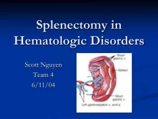
Bone Marrow (BM) Analysis Aspiration usually from posterior iliac crest 0.5-2.0 mL Direct observation of bone marrow activity • Indication • Unexplained anemia and other cytopenias • Unexplained leukocytosis and thrombocytosis • Suspicion of leukemia and myeloproliferative diseases
BM - Cytogenetic Analysis Chromosomal abnormalities - AML, CML, MDS
CML leukocytosis with the presence of precursor cells of the myeloid lineage blood film at 400X