Download
1 / 4
50 likes | 440 Vues
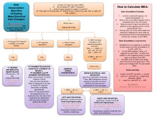
EKG Interpretation Algorithm (including Mean Electrical Axis Changes) ( dxs in light blue= shockable rythms , text in pink = don’t need to know for Mini II). Is there a P wave for every QRS? Are all waves (P, QRS, T) present? Is the P wave Upright in Leads I, II, and III?

E N D
EKG Interpretation Algorithm (including Mean Electrical Axis Changes) (dxs in light blue= shockable rythms, text in pink = don’t need to know for Mini II) • Is there a P wave for every QRS? • Are all waves (P, QRS, T) present? • Is the P wave Upright in Leads I, II, and III? • (IF THE ANS TO EVEN ONE OF THESE IS NO, THEN FOLLOW THE no SIDE OF THE CHART.) • How to Calculate MEA: • Semi-Quantitative Estimate: • Look for a lead with approx. net electrical deviation = 0. • Draw a line on the MEA diagram that is perpendicular to the net 0 lead • Now you know it has to be either the positive or the negative portion of that perpendicular line . • Choose any one of the other leads and draw the 3-segment on each side arc, and whichever half of the perpendicular line the arc crosses, is your MEA. • Semi-Quantitative Long Version: • Establish the net negativity or positivity of each lead on the six limb leads (I, II, II, aVF, aVR, VL) • On the MEA diagram, draw a “3-segment on each side “ on either the positive or the negative portion of each lead, according to the EKG • The MEA must lie within the wedge which has all six arcs spanning it. This gives you a range of 30˚ for your actual MEA. • Quick and Dirty: • Leads I and aVF are both + = normal • Lead I is – and aVF is + = Right Axis Deviation (RAD) • Lead I is + and aVF is - = LAD • Quantitative • (not desc. here b/c requires ruler) YES to ALL = SINUS RHYTHM 4. QRS Complex changes in net electrical deviation from list ? (Net + = Leads I, II, avF, aVL, V5, V6 Net - = aVR, V1) 5. MEA < -90˚ or > +30˚? 1. Prolonged P-R interval? (>.20sec, or 5 small boxes) 2. ST-segment elevation? 3. Other P wave changes? Yes Yes Yes Yes to Any • ST SEGMENT ELEVATION = DIASTOLIC CURRENT OF INJURY = • TP SEGMENT and PR SEGMENT DEPRESSION • dead cells maintain constant negative charge • the only time the whole heart is supposed to be neg is during ST segment (ventricles completely depolarized) • thus, ST seg stays where it’s supposed to be, on isoelectric line, the rest of the segments are depressed with downward deflection. • SINUS RHYTHM • cath lab and/or lytics (cath preferred) • 1st DEGREE • (INCOMPLETE) • HEART BLOCK • PR-interval > .20 sec • SINUS RHYTHM • benign, no urgent intervention required. GENERAL ATRIAL HYPERTROPHY MEAN ELECTRICAL AXIS DEVIATION (see R for calculation methods) **note, MEA deviations can be present in pts with non-sinus rhythms, but they are not reproduced on the next page. 1. MEA < -30˚ to > -90˚? 2. MEA < +90˚ to > +150˚? • LEFT AXIS DEVIATION • pathologic causes include L Ventricular Hypertrophy, Inferior MI, Emphysema, Systemic HTN, Aortic Valve Stensosis • physiologic causes include athletic conditioning • RIGHT AXIS DEVIATION • pathologic causes include R Ventricular Hypertrophy, Lateral MI, Pulmonary HTN, Pulmonary Valve Stenosis, VSD, Tetrology of Fallot • physiologic causes include tall, thin adult, and childhood, high altitude
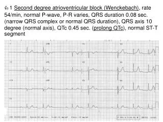
EKG Interpretation Algorithm (not including Mean Electrical Axis Changes) (dxs in light blue= shockable rhythms) • Is there a P wave for every QRS? • Are all waves (P, QRS, T) present? • Is the P wave Upright in Leads I, II, and III? • (IF THE ANS TO EVEN ONE OF THESE IS NO, THEN FOLLOW THE no PAGE OF THE CHART.) 1. Separate P wave and QRS complex rhythm? Y • 3rd DEGREE (COMPLETE)HEART BLOCK aka Atrioventricular Dissociation • P wave has atrial rhythm, QRS wave has Junctional (AV node)or Ventricular (His-Purkinje or Ventricular Myocardium) rhythm • Hallmark: P wave and R wave are said to be “marching out” meaning they follow sep. rhythms, but are still highly regular (p-p and r-r do not change) • Hallmark: P wave found btx QRS and T wave • sometimes: inverted T waves. • Junctional Rhythm = narrow QRS < 3 small boxes • Accelerated Idioventricular Rhythm = widened QRS • tx = pacing, transvenous or transcutaneous • NON-SINUS RYTHM NO to ONE or MORE = NON-SINUS RYTHM Yes 1. Dropped QRS complexes? Yes No 1. Has P Waves? 2. P waves unclear, erratic baseline? Y 1. Total Absence of any waveform pattern? 2. Prolonged PR Interval? No • ATRIAL FIBRILLATION • no clear P waves, still have QRS. no reg. HR • atria contract erratically, causes irregular baseline • not directly fatal, but causes clots • Pulmonary Embolism thrombus formed in atria goes to pulmonary circ and lungs • Coronary or Cerebral Embolism thrombus formed in atrium goes to coronary art. or brain • NON-SINUS RHYTHM No Yes 1. Wide QRS Complex? Yes • 2nd DEGREE • (INCOMPLETE) • HEART BLOCK • MOBITZ type 2 • PR-interval = no Δ • sudden, unpredictable loss of QRS complex. • disease of bundle of His-purkinje system • can be 2:1 or 3:1 (p wave:QRS compl.) • NON-SINUS RHYTHM • can degrade to 3rd deg. heart block • 2nd DEGREE • (INCOMPLETE) • HEART BLOCK • MOBITZ type 1 • aka Wenkebach rhythm • PR-interval > .25 sec • PR-intervals often get progressively longer till you lose one, then it re-sets and then they start to get longer again • AV node is disfctl • NON-SINUS RHYTHM!!! Yes No • VENTRICULAR FIBRILLATION • Highly erratic pattern • fatal if not tx’d • NON- SINUS RHYTHM • VENTRICULAR TACHYCARDIA • 150-250 bpm • frequently due to a re-entrant ventricular pathway caused by scar tissue from previous MI, etc. • SVT • SUPRA –VENTRICULAR TACHYCARDIA • >150 bpm • frequently due to a re-entrant pathway • origin of electrical impulse is in the atria or the AV node
EKG Interpretation Algorithm (including Mean Electrical Axis Changes) ( dxs in light blue= shockable) • Is there a P wave for every QRS? • Are all waves (P, QRS, T) present? • Is the P wave Upright in Leads I, II, and III? • (IF THE ANS TO EVEN ONE OF THESE IS NO, THEN FOLLOW THE no SIDE OF THE CHART.) • How to Calculate MEA: • Semi-Quantitative Estimate: • Look for a lead with approx. net electrical deviation = 0. • Draw a line on the MEA diagram that is perpendicular to the net 0 lead • Now you know it has to be either the positive or the negative portion of that perpendicular line . • Choose any one of the other leads and draw the 3-segment on each side arc, and whichever half of the perpendicular line the arc crosses, is your MEA. • Semi-Quantitative Long Version: • Establish the net negativity or positivity of each lead on the six limb leads (I, II, II, aVF, aVR, VL) • On the MEA diagram, draw a “3-segment on each side “ on either the positive or the negative portion of each lead, according to the EKG • The MEA must lie within the wedge which has all six arcs spanning it. This gives you a range of 30˚ for your actual MEA. • Quick and Dirty: • Leads I and aVF are both + = normal • Lead I is – and aVF is + = Right Axis Deviation (RAD) • Lead I is + and aVF is - = LAD • Quantitative • (not desc. here b/c requires ruler) YES to ALL = SINUS RHYTHM 4. QRS Complex changes in net electrical deviation from list ? (Net + = Leads I, II, avF, aVL, V5, V6 Net - = aVR, V1) 5. MEA < -90˚ or > +30˚? 1. Prolonged P-R interval? (>.20sec, or 5 small boxes) 2. ST-segment elevation? 3. Other P wave changes? Yes Yes Yes Yes to Any • ST SEGMENT ELEVATION = DIASTOLIC CURRENT OF INJURY = • TP SEGMENT and PR SEGMENT DEPRESSION • dead cells maintain constant negative charge • the only time the whole heart is supposed to be neg is during ST segment (ventricles completely depolarized) • thus, ST seg stays where it’s supposed to be, on isoelectric line, the rest of the segments are depressed with downward deflection. • SINUS RHYTHM • cath lab and/or lytics (cath preferred) • 1st DEGREE • (INCOMPLETE) • HEART BLOCK • PR-interval > .20 sec • SINUS RHYTHM • benign, no urgent intervention required. GENERAL ATRIAL HYPERTROPHY MEAN ELECTRICAL AXIS DEVIATION (see R for calculation methods) **note, MEA deviations can be present in pts with non-sinus rhythms, but they are not reproduced on the next page. 1. MEA < -30˚ to > -90˚? 2. MEA < +90˚ to > +150˚? • LEFT AXIS DEVIATION • pathologic causes include L Ventricular Hypertrophy, Inferior MI, Emphysema, Systemic HTN, Aortic Valve Stensosis • physiologic causes include athletic conditioning • RIGHT AXIS DEVIATION • pathologic causes include R Ventricular Hypertrophy, Lateral MI, Pulmonary HTN, Pulmonary Valve Stenosis, VSD, Tetrology of Fallot • physiologic causes include tall, thin adult, and childhood, high altitude
EKG Interpretation Algorithm (not including Mean Electrical Axis Changes) (dxs in light blue= shockable rhythms) • Is there a P wave for every QRS? • Are all waves (P, QRS, T) present? • Is the P wave Upright in Leads I, II, and III? • (IF THE ANS TO EVEN ONE OF THESE IS NO, THEN FOLLOW THE no PAGE OF THE CHART.) 1. Separate P wave and QRS complex rhythm? Y • 3rd DEGREE (COMPLETE)HEART BLOCK aka Atrioventricular Dissociation • P wave has atrial rhythm, QRS wave has Junctional (AV node)or Ventricular (His-Purkinje or Ventricular Myocardium) rhythm • Hallmark: P wave and R wave are said to be “marching out” meaning they follow sep. rhythms, but are still highly regular (p-p and r-r do not change) • Hallmark: P wave found btx QRS and T wave • sometimes: inverted T waves. • Junctional Rhythm = narrow QRS < 3 small boxes • Accelerated Idioventricular Rhythm = widened QRS • tx = pacing, transvenous or transcutaneous • NON-SINUS RYTHM NO to ONE or MORE = NON-SINUS RYTHM Yes 1. Dropped QRS complexes? Yes No 1. Has P Waves? 2. P waves unclear, erratic baseline? Y 1. Total Absence of any waveform pattern? 2. Prolonged PR Interval? No • ATRIAL FIBRILLATION • no clear P waves, still have QRS. no reg. HR • atria contract erratically, causes irregular baseline • not directly fatal, but causes clots • Pulmonary Embolism thrombus formed in atria goes to pulmonary circ and lungs • Coronary or Cerebral Embolism thrombus formed in atrium goes to coronary art. or brain • NON-SINUS RHYTHM No Yes 1. Wide QRS Complex? Yes • 2nd DEGREE • (INCOMPLETE) • HEART BLOCK • MOBITZ type 2 • PR-interval = no Δ • sudden, unpredictable loss of QRS complex. • disease of bundle of His-purkinje system • can be 2:1 or 3:1 (p wave:QRS compl.) • NON-SINUS RHYTHM • can degrade to 3rd deg. heart block • 2nd DEGREE • (INCOMPLETE) • HEART BLOCK • MOBITZ type 1 • aka Wenkebach rhythm • PR-interval > .25 sec • PR-intervals often get progressively longer till you lose one, then it re-sets and then they start to get longer again • AV node is disfctl • NON-SINUS RHYTHM!!! Yes No • VENTRICULAR FIBRILLATION • Highly erratic pattern • fatal if not tx’d • NON- SINUS RHYTHM • VENTRICULAR TACHYCARDIA • 150-250 bpm • frequently due to a re-entrant ventricular pathway caused by scar tissue from previous MI, etc. • SVT • SUPRA –VENTRICULAR TACHYCARDIA • >150 bpm • frequently due to a re-entrant pathway • origin of electrical impulse is in the atria or the AV node