Download
1 / 69
700 likes | 1.2k Vues
The Immune System and Mechanisms of Defense. 0. 9. Overview of the Body’s Defense Mechanisms. Defense mechanisms include Barriers to entry of pathogens (disease-producing microorganisms) Skin, stomach acid, tears, vomiting Nonspecific defense mechanisms Phagocytosis, inflammation

E N D
Overview of the Body’s Defense Mechanisms Defense mechanisms include Barriers to entry of pathogens (disease-producing microorganisms) Skin, stomach acid, tears, vomiting Nonspecific defense mechanisms Phagocytosis, inflammation Specific defense mechanisms Immune response enables body to recognize and remove specific bacteria, other foreign cells, viruses Antibodies T cells
Pathogens Cause Disease Disease-causing agents include Living organisms Bacteria: unicellular prokaryotes Fungi: unicellular and multicellular eukaryotes Parasites: unicellular and multicellular eukaryotes Nonliving infectious “particles” Viruses Prions
Figure 9.1 SEM ( 12,000) of Campylobacter jejuni, a spiral-shaped bacterium that causes food poisoning. SEM ( 2,000) of Streptococcus, a spherical bacterium that causes sore throats. SEM ( 5,600) of Escherichia coli, a common intestinal bacteria that is usually harmless.
Figure 9.2 Ribosomes DNA Cell wall Relative size of a bacterium Plasma membrane Mitochondrion Bacterial cell. Bacteria have a single strand of DNA and free-floating small ribosomes within their cytoplasm. Their plasma membrane is surrounded by a rigid cell wall. Nucleus Golgi apparatus RNA Plasma membrane Relative size of a virus DNA Protein coat Endoplasmic reticulum Viruses. Viruses consist of a protein coat surrounding either RNA or DNA. Eukaryotic cell. Eukaryotic cells have a membrane-bound nucleus and well-defined membrane-bound organelles.
Bacteria: Single-Celled Living Organisms Characteristics Prokaryotic (lack a membrane-enclosed nucleus and membranous organelles) Single-celled Use a variety of resources for growth and reproduction Infections Pneumonia, tonsillitis, tuberculosis, botulism, toxic shock syndrome, syphilis, Lyme disease, etc. Generally treated with antibiotics
Viruses: Tiny Infectious Agents Extremely small, much smaller than bacteria Living? Open to debate Unable to reproduce outside of a host cell No metabolic activity Structure Contain DNA or RNA, not both Nucleic acid is surrounded by a protein coat Diseases caused by viruses: AIDS, hepatitis, encephalitis, rabies, influenza, colds, warts, chicken pox
Animation: Structure and Reproduction of Viruses Right-click and select Play
Prions: Infectious Proteins Infectious proteins Normal brain proteins that are not folded correctly The mis-folding becomes self-propagating, filling and disabling the cell with protein debris Resist cooking, freezing, drying Diseases Bovine spongiform encephalitis (BSE, “mad cow disease”) Variant Creutzfeldt-Jakob disease (vCJD)
Transmissibility, Mode of Transmission, and Virulence Determine Health Risk Transmissibility How easily a pathogen is passed from person to person Mode of transmission Respiratory, fecal–oral, body fluids, direct contact Virulence How much damage is caused by the infection
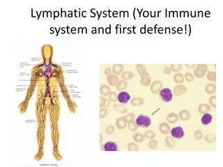
The Lymphatic System Defends the Body Functions Maintenance of blood volume in cardiovascular system Transport of fats and fat-soluble material from digestive system to cardiovascular system Filtration of foreign material to defend against infection
Figure 9.3 Nasal passages Thymus gland Adenoids Tongue Tonsils Heart Trachea Tonsils protect the throat. Lymphocytes mature in thymus. Lymph flow Blood flow Red pulp Lymph node White pulp Lymph vessels Spleen Macrophages cleanse lymph; lymphocytes activate defense mechanisms. Spleen removes damaged blood cells and microorganisms from blood. Blood capillary Lymphatic vessels transport fluid, bacteria, and viruses. Lymphatic capillary Cells
Lymphatic Vessels Transport Lymph Lymphatic vessels transport lymph Begin as blind-ended lymphatic capillaries Network of vessels, similar to veins Eventually drain into cardiovascular system through right lymphatic duct and thoracic duct Lymph is a milky fluid containing: White blood cells Proteins Fats Occasionally bacteria and viruses
Lymph Nodes Cleanse the Lymph Lymph nodes are located at intervals along lymphatic vessels Nodes remove microorganisms, debris, and abnormal cells from lymph Small, 1mm to 2.5 mm in size Nodes are composed of connective tissue, macrophages, and lymphoctyes Nodes act as filters, cleansing the lymph as it passes through them
The Spleen Cleanses the Blood Largest lymphatic organ Located in upper left abdominal cavity Two regions of spleen: Red pulp Removes old and damaged red blood cells Temporary blood storage White pulp Contains lymphocytes, searching for pathogens Diseases that cause spleen enlargement Infectious mononucleosis, leukemia Spleen can be removed with minor medical impact
Thymus Gland Hormones Cause T Lymphocytes to Mature Thymus gland Located behind sternum, above heart Site of maturation of T cells (T lymphocytes) Secretes two hormones that control T cell development: thymosin and thymopoietin Largest, most active during childhood Atrophies with aging Tonsils Filter food and air entering the throat Adenoids Filter air, back of nasal passages
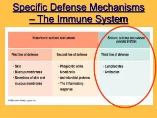
Keeping Pathogens Out: The First Line of Defense Skin—an effective deterrent Tears and saliva—contain lysozyme (antibacterial enzyme) Ear wax—entraps microorganisms Mucus—entraps microorganisms Stomach—highly acidic, inhibits microorganisms Vagina—slightly acidic, inhibits some microorganisms Vomiting, urination, and defecation—remove microorganisms Resident bacteria—outcompete pathogens
Nonspecific Defenses: Second Line of Defense Phagocytic cells: white blood cells that surround and engulf invading bacteria Neutrophils, macrophages, eosinophils Inflammation Redness, warmth, swelling, pain Natural killer cells: a type of lymphocyte that attacks tumor cells and virus-infected cells Complement proteins: plasma proteins that invade bacteria when activated Interferons: antiviral proteins Fever response
Figure 9.6 1 Phagocyte approaches and captures bacterium. 2 Phagocyte surrounds bacterium. Bacterium Vesicle 3 Bacterium becomes enclosed in vesicle. Lysosome 4 Vesicle fuses with lysosomes. An electron micrograph of a macrophage (blue) capturing several bacteria (pink). 5 Lysosomal enzymes digest bacterium. Cytoplasm of phagocyte 6 Wastes and debris are discarded. Steps in the process.
Figure 9.7 Phagocyte Site of injury Mast cell Complement protein Bacteria Histamine 1 2 3 Complement proteins from plasma diffuse out of leaky capillaries. They mark the bacteria for destruction and sometimes kill them. Attracted by histamine and other chemicals, phagocytes squeeze through the leaky capillary walls and begin attacking and engulfing bacteria and debris. Damaged cells and mast cells in the area release histamine and other substances. Histamine dilates blood vessels and makes them leaky.
Animation: The Inflammation Response Right-click and select Play
Figure 9.8 1 2 3 The bacterium swells and eventually bursts. Activated complement proteins form complexes of proteins that create holes in the bacterial cell wall. Water and salts diffuse into the bacterium through the holes. Water and salts Complement proteins Bacterium Cell wall of bacteria Photomicrograph of an intact bacterium A bacterium after lysis by activated complement proteins.
Specific Defense Mechanisms: The Third Line of Defense The immune response Characteristics Recognizes and targets specific pathogens and foreign substances Has “memory”—“remembers” initial exposure and responds more quickly and aggressively on subsequent exposures Able to distinguish between “Self” cells and foreign, “non-self” invaders Healthy cells and abnormal (tumor) cells
The Immune System Targets Antigens Antigen: any substance that triggers an immune response Usually protein or polysaccharide on outer surface of invading cell or virus MHC (major histocompatibility complex) proteins Self-antigens that are on human cell surfaces enabling recognition of “self” Enable immune system to distinguish “self” from “non-self”
Lymphocytes Are Central to Specific Defenses B lymphocytes: Antibody-mediated immunity Antibodies: proteins made by B lymphocytes that bind with and neutralize specific antigens Active against viruses, bacteria, and soluble foreign molecules T lymphocytes: Cell-mediated immunity Directly attack foreign cells Coordinate the immune response Active against parasites, viruses, fungi, intracellular bacteria, cancer cells, cells with “non-self” MHC
B cells: Antibody-Mediated Immunity B cells activated when they recognize an antigen Divide into two cell types Memory cells—store information for future immune responses Plasma cells—actively secrete antibodies, which will bind to antigen
Figure 9.9 Bacterium with surface antigens Mature inactive B cells specific for different antigens, found in lymphatic tissue Binding, activation Clone formation Plasma cells Memory cells Antibodies Memory cells store information until the next exposure to the same antigen. Plasma cells secrete antibodies into circulation.
Figure 9.10 When antibodies encounter a pathogen with the right surface antigen, they bind to it, forming an antigen-antibody complex. Pathogens Antibody Antigen-antibody complex Some antibodies cause pathogens to agglutinate (clump together). The formation of an antibody-antigen complex marks the pathogen for attack by phagocytes or complement proteins.
The Five Classes of Antibodies Antibodies also known as immunoglobulins (Ig) Classes of antibodies IgG: most prevalent in the blood IgM: first antibody produced in an immune response IgA: found in body secretions, including breast milk IgD: function is unclear IgE: plays a key role in allergic responses
Figure 9.11 Antigen Antigen- binding site Variable regions Light chain Heavy chain Constant regions
T Cells: Cell-Mediated Immunity T cells Originate from stem cells in the bone marrow Mature in the thymus Types of T cells CD4 T cells Helper T cells and Memory T cells CD8 T cells Cytotoxic T cell
T Cells: Cell-Mediated Immunity T cells must be presented with antigen by antigen-presenting cells (APCs) APCs include Macrophages B cells
Figure 9.12 Antigen Major histocompatibility complex protein (MHC) Pathogen 1 The macrophage engulfs a pathogen. Lysosome 2 Vesicle with MHC molecules Lysosomes partially digest the pathogen. 3 A vesicle containing MHC molecules binds to the digestive vesicle. 4 The MHC molecules and a fragment of the antigen form an antigen-MHC complex. 5 The antigen-MHC complex is displayed on the surface of the cell when the vesicle fuses with the cell membrane and releases its digestive products. Antigen-MHC complex
T Cells: Cell-Mediated Immunity Helper T cells Secrete cytokines, which stimulate other immune system cells Play a key role in directing the immune response Are targets of HIV infection Cytotoxic T cells Directly attack and destroy abnormal (tumor or viral-infected) cells and foreign cells Memory T cells Reactivate during later exposures
Figure 9.13 MHC molecule Antigen-presenting cell (APC) CD4 receptor Antigen fragment Inactive helper T cell 1 Activation Memory T cells 2 Clonal expansion 3 Cytokine production
Figure 9.14 MHC molecule Antigen-presenting cell (APC) CD8 receptor Antigen fragment Inactive cytotoxic T cell 1 Activation Memory T cells 2 Clonal expansion 3 Attack on target cell
Figure 9.15 Cytotoxic T cell Target cell Cytotoxic T cells (blue) attaching to a target cell (pink). Cytotoxic T cell Vesicle Perforin Granzyme Cytotoxic T cell membrane Intercellular space 3 Completed pore; granzyme passing through 2 1 Perforin pore partially assembled Intact target cell membrane Target cell How cytotoxic T cells kill a target cell.
Immune Memory Creates Immunity Primary immune response Occurs on first exposure to antigen Characteristics Lag time of 3–6 days for antibody production Peak at 10–12 days Secondary immune response Occurs on second and subsequent exposure to antigen Characteristics Lag time in hours Peak in days Much more antibody produced
Figure 9.16 Secondary immune response Primary immune response 100 10 Antibody concentration (units/ml) 1 0.1 0 7 14 21 28 0 7 14 21 28 35 42 1st exposure 2nd exposure Time (days after exposure)
Medical Assistance in the War Against Pathogens Immunization A strategy for causing the body to develop immunity to a specific pathogen Active immunization Intentionally expose individual to a form of the antigen that doesn’t produce disease (vaccine) Also known as vaccination Passive immunization Administer protective antibodies to an individual
Medical Assistance in the War Against Pathogens Monoclonal antibodies Specific antibodies produced in the laboratory by a hybrid B cell clone Commercial applications of monoclonal antibodies Home pregnancy tests Prostate cancer screening test Diagnostic testing for hepatitis, influenza, HIV
Figure 9.17 1 Immunize mouse with antigen. 2 Extract B cells from the mouse’s spleen. Myeloma (cancer) cells 3 Fuse antibody-producing B cells with cancer cells to produce fast-growing cells. Hybridoma cell 4 Select cells that produce the desired antibody. Hybridoma cells multiply in culture and produce antibodies 5 Clone antibody-producing hybridoma cells. 7 Extract the antibodies. 6 Grow large numbers of the cells in culture.
Medical Assistance in the War Against Pathogens Antibiotics combat bacteria Antibiotics kill bacteria or inhibit their growth Antibiotics are selectively toxic for bacteria by targeting features of bacterial cells that are different from eukaryotic cells Antibiotics are not effective against viruses
Tissue Rejection: A Medical Challenge Tissue rejection May occur following tissue or organ transplant if recipient’s immune system attacks the transplanted tissue/organ To minimize risk of rejection Must match ABO and other blood group antigens and MHC antigens 75% MHC match is essential MHC antigens allow body to distinguish “self” from “nonself” Immunosuppressive drugs—prevent patient’s immune system from attacking transplanted tissue
Inappropriate Immune System Activity Causes Problems Allergies are hypersensitivity reactions Inappropriate response to an allergen Allergen: any substance (antigen) that causes an allergic reaction (not a pathogen, but the body reacts as though it is a pathogen) Examples of allergens Pollen Bee venom Foods (nuts, seafood) Oil from poison ivy plan
Inappropriate Immune System Activity Causes Problems Allergies (cont’d) Excessive inflammatory response mediated by IgE Basophils and mast cells Histamine Reactions may be localized or systemic Localized: affect only the area exposed Systemic: affect several organ systems Anaphylactic shock: severe life-threatening systemic reaction (difficulty breathing, circulatory collapse, drop in blood pressure)
Inappropriate Immune System Activity Causes Problems Allergies (cont’d) Treatment of allergies Antihistamines—treatment of mild to moderate reactions Epinephrine injection—treatment of anaphylactic shock Allergy shots