PET Imaging for Early Alzheimer's Disease Detection: A Ray of Hope!
Explore the grave threat of Alzheimer's disease, its alarming statistics, and cognitive impairments. Learn about the challenges in diagnosing AD and the vital need for early detection. Discover how PET imaging can provide a ray of hope by aiding in the preclinical diagnosis and predicting the risk of developing AD. Understand the clinical edge PET scans offer over other imaging techniques and their common applications in Alzheimer's disease and other disorders.

PET Imaging for Early Alzheimer's Disease Detection: A Ray of Hope!
E N D
Presentation Transcript
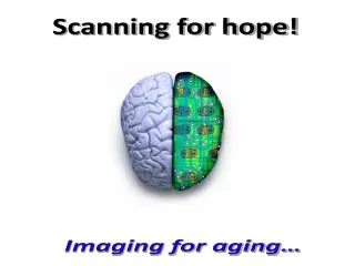
Scanning for hope! Imaging for aging…
Alzheimer’s disease – A grave threat Or… a threat that leads to the grave?
Alzheimer’s disease Statistics are alarming • There are currently 26 million people worldwide with Alzheimer’s disease. This figure is projected to grow to more than 106 million people by 2050. • More than 5 million Americans are believed to have Alzheimer’s disease and by 2050, the number could increase to 15 million. • Approximately 350,000 new cases of Alzheimer's disease are diagnosed each year. • Alzheimer’s disease is the most common form of dementia (represents about 60% of the cases of dementia in the elderly). • Death from Alzheimer’s disease is often underreported or misdiagnosed.
Alzheimer’s disease Understanding the disorder “Alzheimer's disease (AD) is chronic, progressive, age-related, non-reversible and incurable (to date) brain disorder that develops over a period of years and ultimately leads to a severe loss of mental function. Alzheimer's disease (AD) gradually results in cognitive impairments that leave patients completely dependent on others.”
Diagnosis of Alzheimer’s disease The problem “Until today, Alzheimer’s disease cannot be definitively diagnosed until after death, when a brain autopsy is performed on a patient and evidence of beta-amyloid plaque deposits and NFTs are found.”
Diagnosis of Alzheimer’s disease The challenge “In AD, the development of pathology in the brain is thought to precede cognitive symptoms and, hence, diagnosis of the disease, by many years. Therefore, preclinical diagnosis of Alzheimer’s disease is one of the major challenges for the prevention of AD.”
Diagnosis of Alzheimer’s disease The options Structural imaging Functional imaging X Ray PET CT SPECT MRI Dce MRI
Diagnosis of Alzheimer’s disease MRIs & CT scans have limitations “CT scans and MRIs only detect structural or anatomical changes in the brain and detection of subtle pathophysiological change in the brain at asymptomatic stages is not possible with MRI and CT scan especially when there is no evidence of anatomic changes.”
Diagnosis of Alzheimer’s disease Early detection can be vital “Early diagnosis of Alzheimer’s disease might enable effective treatment before so many brain cells are lost through the disease. Therefore, it is important to identify non-demented individuals in the preclinical stage of AD, who are at very high risk for developing dementia.”
Diagnosis of Alzheimer’s disease A ray of hope! “Brain imaging, PET amyloid imaging in particular, when combined with the specific biomarkers, greatly increases one’s ability to predict who is at risk of developing AD several years before disease onset. It is now widely believed that β-Amyloid (Aβ) imaging has great potential to aid in the diagnosis of Alzheimer disease and the development of related therapeutics.”
Amyloid imaging via Positron emission tomography (PET) A possible answer to the challenge? “Positron emission tomography, also called PET imaging or a PET scan or nuclear medicine scan, is a type of nuclear medicine imaging which produces 3-dimensional, color images of the functional processes within the human body or brain.”
Amyloid imaging via Positron emission tomography (PET) Defining PET “Positron emission tomography (PET) is a method of medical imaging which allows displaying metabolic activity in a slice of the body by means of detecting radiation, emitted from a radio-isotope injected into the patient’s body.”
Positron emission tomography (PET) What makes PET so unique? Today, a whole-body PET scan provides information about the body’s chemistry and cell function (metabolism) rather than pictures of the body’s anatomy or structure as shown by X-ray, ultrasound, CT scans, or MRI. As a result, PET scans may reveal abnormalities or tumors that would otherwise go undetected.
Positron emission tomography (PET) Time line of PET scanning at a glance • 1975 the first commercial PET scanner was introduced • 70s and 80s PET was mainly used for research • 1990s being used in clinics regularly
Positron emission tomography (PET) Basic working principle “PET is an imaging technique that can quantify increases in nerve cell activity in selective regions of the specific part of the body such as brain. PET scans use a small amount of a radioactive drug, or tracer, to show differences between healthy tissue and diseased tissue.”
Positron emission tomography (PET) The clinical edge over other imaging techniques (2) “As the origins of many diseases are biochemical in nature, these functional changes often predate or exceed structural change in tissue or organs. PET enables physicians to assess chemical or physiological changes related to metabolism.”
Positron emission tomography (PET) Common clinical / diagnostic applications Alzheimer's disease Mobility disorders Neoplasms PET uses Lung pathologies Neurological disorders e.g. epilepsy Visual cortex
Positron emission tomography (PET) Stages in PET imaging 1 Radiotracer injection 2 Positron emission & annihilation 3 Image production (reconstruction)
Stages in PET imaging Defining the radiotracer “PET scanning uses artificial radioactive tracers and radionuclides. Their lifetime is usually rather short, thus they need to be produced on site. PET tracers mimic the natural sugars, water, proteins, and oxygen found in our bodies. These tracers are injected into a patient and collect in various tissues and organs.”
Stages in PET imaging 1- Radiotracer injection “A metabolically active tracer, a biologic molecule that carries with it a positron emitting isotope is inserted in the body. Over a few minutes the isotope accumulates in an area of the body for which the molecule has an affinity. i.e. glucose labeled with 11C or glucose analogue labeled with 18F, accumulates in the brain or tumors, where glucose is used as the primary source of energy.”
Stages in PET imaging 2- Positron emission & annihilation “The radioactive nuclei then decay by positron emission as a nuclear proton changes into a positive electron and a neutron. The ejected positron combines with an electron instantaneously, and these 2 particles undergo the process of annihilation. The energy associated with the masses of the positron and electron is divided equally between 2 photons which fly away from one another at 1800 angle. Each photon has an energy of 511 keV. These high energy gamma rays emerge from the body in opposite directions and recorded simultaneously by pair of detectors. “
Stages in PET imaging 3- Photo / image generation (reconstruction) “After 100,000 or more annihilation events are detected, the distribution of the positron-emitting tracer is calculated by tomographic reconstruction procedures. PET reconstructs a 2 dimensional image of cellular biological activities from the one dimensional projections seen at different angles. 3-D reconstructions can be done using 2D projections from multiple angles.”
Positron emission tomography (PET) Illustrative example of working
PET – Amyloid imaging Frequently used radiotracers
PET – Amyloid imaging Limitations of common radiotracers • PIB (Pittsburgh Compound B ) • Short (20.4-min) radioactive half-life • Higher dose is required
PET – Amyloid imaging Limitations of common radiotracers • FDG (2-[18F] fluoro-2-deoxy-D-Glucose) • In very early disease, the scans may be normal or equivocal. • accuracy declines in the very elderly patient.
PET – Amyloid imaging 18F-BAY94-9172 – A promising PET tracer • 18F-BAY94-9172 • Significantly longer half life ( 109.4 m) • Has shown high affinity and specificity for Aβ in vitro and binding to Amyloid plaques. • Can distinguish subjects with AD from healthy elderly subjects and subjects with frontotemporal dementia
PET – Amyloid imaging Features versus benefits
Some in-demand examples of health feature writing Correct diagnosis Confirmed, accurate diagnosis Early diagnosis PET-Amyloid imaging Fast diagnosis Economical Safe & compliant
PET-Amyloid imaging A true game changer “The radioactive nuclei then decay by positron emission as a nuclear positron and electron is divided equally between 2 photons which fly away from one another at 1800 angle. Each photon has an energy of 511 keV. These high energy gamma rays emerge from the body in opposite directions and recorded simultaneously by pair of detectors.”