Download
1 / 23
240 likes | 493 Vues
PLAQUE FORMATION AND BIOCHMICAL MARKERS OF ISCHEMIA. Dr Azra Parveen Senior Registrar Medicine. Acute Myocardial Infarction. Acute myocardial infarction is the rapid development of myocardial necrosis caused by a critical imbalance between the oxygen supply

E N D
PLAQUE FORMATION AND BIOCHMICAL MARKERS OF ISCHEMIA Dr Azra Parveen Senior Registrar Medicine
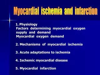
Acute Myocardial Infarction Acute myocardial infarction is the rapid development of myocardial necrosis caused by a critical imbalance between the oxygen supply and demand of the myocardium. • It is an irreversible myocardial injury from prolonged ischemia. • Accurate and early diagnosis is important in minimizing cellular damage and, consequently, in obtaining a successful outcome for the patient
Plaque formation • Risk Factors: Hyperlipiemia-LDL Diabetes Hypertension Smoking Obesity
Initiation of atherosclerosis Expression of adhesion molecules that allows the leukocytes to stick to the arterial wall. Early lesion- Fatty streak Upon entry into the arterial wall blood monocytes begin to scavenge lipids and become foam cells.
Stable atherosclerotic plaque Foam cells release further cytokines and effector molecules that stimulate smooth muscle cell migration from the arterial intima into the media, as well as smooth cell proliferation. Smooth muscle cells together with lipd core and matrix form a stable atherosclerotic plaque that will remain asymptomatic until it becomes large enough to obstruct arterial flow.
Vulnerable plaque Inflammatory cell infiltrate, smooth muscle cell death through apoptosis and matrix degradation by matrix metalloproteinases generate a plaque with a thin fibrous cap and lipid-rich necrotic core. It is called a vulnerable plaque and it can rupture. Plaque rupture will expose its contents to blood and trigger platelet aggregation and thrombosis. It will result in partial or complete obstruction of the blood vessel leading to ischemia or infarction
Cardiac Markers Markers of cardiac myocyte necrosis: Myoglobin CK Ck-MB Troponin I & T
Myoglobin • Myoglobin is an iron and oxygen binding protein found in muscle tissue • It is only found in the blood stream when it is released following muscle injury • It is a sensitive marker for mucle injury making it a potential marker for myocardial infarction • However elevated myoglobin has low specificity for the diagnosis of myocardial infarction and therefore is not the preferred test
Creatinekinase • Cytoplasmic CK is a dimer, composed of M and/or B subunits, which associate forming CK-MM, CK-MB and CK-BB isoenzymes. • CK-MM is the main isoenzyme found in striated muscle • CK-MB is found mainly in cardiac muscle • CK-BB is the predominant isoenzyme found in brain
Serum total CK activity and CK-MB concentration rise in parallel following myocardial injury, starting to increase 4± 6 h after injury, reaching peak serum concentrations after 12±24 h and returning to baseline after 48±72 h. Serum CK-MB is considerably more specific for myocardial damage than is serum total CK, which may be elevated in many conditions where skeletal muscle is damaged. Consequently, CK should not be used for the diagnosis of myocardial injury unless used in combination with other more specific cardiac markers.
Total CK can be elevated • • IM injection • • Traumatic damage to skeletal muscle • • Hypothermia • • Exercise • • Intoxication • • Dose-related side effect in statin treatment
CK-MB FRACTION • CK-MB fraction= CKMB /total CK • Rationale for using CK-MB fraction • CK-MB fraction greater than 2.5% is suggestive of myocardial injury
Troponins Troponin is a regulatory complex of 3 protein subunits located on the thin filament of the myocardial contractile apparatus. Its function is the regulation of striated and cardiac muscle contraction. The complex regulates the calcium-modulated interaction between actin and myosin on the thin filament. • Troponin C (18 kd) • • Calcium-binding subunit • • No cardiac specificity • • Troponin I (26.5 kd) • • Actomyosin-ATP-inhibiting • subunit • • Cardiac-specific form • • Troponin T (39 kd) • • Anchors troponin complex • to theTropomyosin strand
In the absence of calcium ions, tropomyosin blocks access to the mysosin binding site of actin. When calcium binds to troponin, the positions of troponin and tropomyosin are altered on the thin filament and myosin then has access to its binding site on actin.. • When the calcium level decreases, troponin locks tropomyosin in the blocking position and the thin filament slides back to the resting state.
Tissue Specificity of TroponinSubunits • Troponin C is the same in all muscle tissues • Troponins I and T have cardiac-specific forms, cTnI and cTnT • cTnI and cTnT remain elevated for 10 to 14 days
Troponin Release Kinetics • Detectable in blood 4-12 h, similar to CKMB • Peaks 12-38 h • Remains elevated for 10-14 days