Download
1 / 1
10 likes | 226 Vues
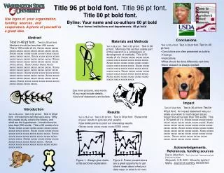
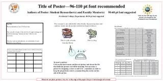
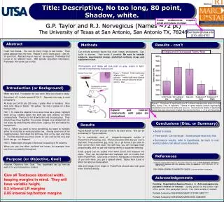
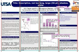
Title: Descriptive, not too long, large (96 pt), shadow, white. Some conferences require email and phone number. G.P. Taylor and R.J. Norvegicus (Names ~72 pt) The University of Texas at San Antonio, San Antonio TX, 78249. Gail.taylor@utsa.edu (210) 458-5761. Methods. Results - con’t.

E N D
Title: Descriptive, not too long, large (96 pt), shadow, white Some conferences require email and phone number. G.P. Taylor and R.J. Norvegicus (Names ~72 pt) The University of Texas at San Antonio, San Antonio TX, 78249 Gail.taylor@utsa.edu (210) 458-5761 Methods Results - con’t Abstract Conclusions (Disc. or Summary) Can include summary figure; flow chart, image, photographs. Can bullet to shorten. Few words as possible. sure to include subjects, experimental design, statistical methods, drugs and equipment used Insert Abstract here. May not be required. Preferably that turned in for abstract book. Will provide redundant information. Preferably around 300ish words (as conference requires). • Large. • Bullet to stress. • Few words. Some people read only this. • Tie back to real world problem brought up in introduction • VERY briefly summarize results. • How did your hypothesis work out? • Why was your finding important? • Future Directions Compound X decreased Tumor size Photographs and tables will look best on gray, cream or light-colored complimentary background. Introduction (Background) Figure 1. Protocol. Tumor cells were obtained from ______________ and cultured. 1 million cells were injected IP into an immune deficient mouse (which). After __________. There should be more words here as well, so that you don’t end up with a horrible white gap in your poster. Enlarge/resize until all space is filled up very cleanly and neatly. Expand and contract components until gaps are minimalized Make very brief. Foundation for your work. Why you chose to study. Equivalent of 1 double spaced 81/2x11. Separate into easy to read paragraphs. Hint 1. To make the objects in one area move as a group, highlight them all by holding down the shift key and clicking on them consecutively. Then go to the draw button and choose group. They should now move together. If you have images on a background, do not resize by stretching the whole item; ungroup first and resize the background. Before you turn it in for printing, please ungroup everything. Hint 2. When you want to move something, but want to maintain either its horizontal or vertical position (ex., moving down one of the heading boxes), hold down the shift key, click on the item and drag it. The first direction you move (horiz. or vert.) will be the only way that it can go…no diagonal. All body text 26-30 pts (28 here). I prefer Ariel or Verdana. Nice dark color (blue or black). No yellow. No red or yellow on a blue background. When you use one effect (outlined text boxes, for example) then use them consistently throughout. References • Quigley HA, Nickells RW, Kerrigan LA, Pease ME, Thibault DJ, Zack DJ. Retinal ganglion cell death in experimental glaucoma and after axotomy occurs by apoptosis. Invest Ophthalmol Vis Sci 1995;36:774-86. , Figure 2. Compound X significantly (p<0.01) shrank osteosarcoma tumors in nude mice, during 1 mo. of treatment. Tumors in carrier-injected controls significantly increased in size over the same time (p<0.01). No behavioral effects or significant weight loss were observed in treated mice during this time. n = 5 at all time points. Results Figure Based. Put words to explain figures so that they can stand alone. Try using figures/graphs most, because they convey the most information. If you use a table, you must add in words near it to explain it and its significance. Manipulate images/photographs outside of PowerPoint (Photoshop, etc). Try not to resize within PowerPoint, because of possible printing problems. They should be at 240 dots per inch (dpi, pixels) and of the correct size. They should be .jpg, .bmp, .tif, or .gif format. Excel graphs can be copied from within Excel and dropped into place. They can be stretched and reshaped with no trouble from within PowerPoint. Click once on them them to manipulate or format them (if you click twice, you get a spread sheet). If you click on their corner then hold down the shift key, you will increase them proportionally, and not get odd-looking skinny or squashed lettering. Tables from Excel or Word can also be manipulated. Time Course of Tumor Reduction Are required - Standard Format. Can make smaller if needed In this column, items are positioned horizontally, 27.5” from the top left corner Purpose (Objective, Aim, Goal) Purpose, Objective, Aim, Goal: What are you going to do? What is your hypothesis. Put it here, or in your Intro but bolded, or you can even create another heading for hypothesis. Acknowledgements Funding Organization/Agency/Source & Acknowledgements, possible conflicts of interest - usually placed at the bottom right of the poster. One paragraph (short). Can make smaller if needed. Partially funded by NIH/NIGMS MARC U*STAR GM07717 Partially funded by NIH/NIGMS MBRS-RISE GM6065 All Textboxes 12” wide, variable ht. 0.2 internal LR margins0.05 internal top/bottom margins In this column, items are positioned horizontally, 1.0” from the top left corner In this column, items are positioned horizontally, 40.75” from the top left corner In this column, items are positioned horizontally, 14.25” from the top left corner Figure 3. Progressive shrinkage of tumor size over 1 mo. with treatment of high dose compound X ((p<0.01). Tumors in carrier-injected controls significantly (p<0.01) increased in size over the same time. n = 5 at all time points.