Download
1 / 52
560 likes | 700 Vues
Explore the fascinating world of cells with this comprehensive introduction. From the basics of cellular life to the evolution of metabolism and the history of cell studies, uncover the secrets of this fundamental unit of life. Learn about the classification of cells, the first cell on Earth, and the significant figures in cell biology history. Dive into the cell theory and discover how artists and scientists perceive cells. With insights on microscopes and their role in studying cells, embark on an enlightening tour of the cell.

E N D
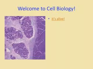
Introduction to cell biology A Tour of the Cell F. Rajaei, PhD Qazvin University of Medical Sciences
سرفصل عناوین • منابع: • زیست شناسی سلولی وملکولی تالیف دکتر احمد مجد • 2-The cell (Albert) • 3-Lodish Molecular Cell Biology (Harvey Lodish, et al) • 4- Medical Cell Biology (Steven R. Goodman)
What is exactly is life? • From the Biological perspective- Life is described with ALL the following 4 properties (at this time) • CELLULAR: Firstly, every living thing is cellular • it is either a single-celled creature (unicellular - bacterium, brewers yeast, amoeba) • or a creature composed of many cells (muticellular - toadstool, frog, plant, man) • PROPERGATE: Living things reproduce themselves • Either individually (asexual reproduction) • In sexual pairs (sexual reproduction • METABOLIZE: Life uses processes collectively called metabolism to convert materials and energy for its needs • EVOLUTION: Life undergoes evolution to different forms
LIFE • -There is no hard and fast definition of life! • -Scientists are manipulating life at this time! • -New life is being created in test tubes! • -NASA is looking for new life forms at this time
What is living? • Animals • Plants • Fungi • Bacteria • Viruses • Prions • Atoms
What is living? • Animals - yes • Plants - yes • Fungi - yes • Bacteria - yes • VIRUSES - no • PRIONS - no • ATOMS - no
Classification of cells • -Two main classes of cells - so far! • Nucleus - Do they have or not have? • - Prokaryotic cells - All Bacteria & Archaea - no nucleus • Eukaryotic cells - Plants, animals, fungi, and protists - all have a nucleus • -Prokaryotes also lack organelles or cytoskeleton • -Otherwise, they function very much the same.
The First Cell • -Earth formed @ 4.5 billion years ago • -Life emerged @ 3.8 billion years ago • -No or little oxygen • -CO2 and N2; H2, H2S and CO • -Add heat and water • -Organic molecules • -LIFE! ????????
The First Cell • -Proteins and RNA were thought to be the first macromolecules produced • -Only RNA capable of directing its self-replication, acting as both template and catalyst • -“RNA World” existed for a period of time • -The FIRST CELL is thought to have arisen when RNA became enclosed in a phospholipid membrane, and was able to self-replicate
Evolution of Metabolism • -The early Earth lacked O2 • -Early Cells took food and energy by absorption • -Glycolysis - glucose to lactic acid • -Photosynthesis - sunlight, water, CO2 to Glucose and O2 • -Oxidative metabolism - Glucose and O2 to CO2 and water • -ATP is generated • -All Cells use ATP for energy to drive their cellular machinery
Cell History • Cytology- study of cells • 1665 English Scientist Robert Hooke • Used a microscope to examine cork (plant) • Hooke called what he saw "Cells"
History of The Cell • -Looked at a thin slice of cork under a microscope • Observed hollow, tiny, room like structures • -Only saw the cell wall because cork cells are not alive • -Called “cells” because they resemble rooms Robert Hooke http://commons.wikimedia.org/wiki/File:Robert_Hooke_portrait.jpg http://www.britannica.com/EBchecked/topic-art/380414/99713/Robert-Hookes-drawings-of-the-cellular-structure-of-cork-and
Cell History • Robert Brown • discovered the nucleus in 1833. • Matthias Schleiden • German Botanist Matthias Schleiden • 1838 • ALL PLANTS "ARE COMPOSED OF CELLS". • Theodor Schwann • Also in 1838, • discovered that animals were made of cells
Cell History • -Rudolf Virchow • 1855, German Physician • " THAT CELLS ONLY COME FROM OTHER CELLS". • -His statement debunked • "Theory of Spontaneous Generation"
Cell Theory • The COMBINED work of Schleiden, Schwann, and Virchow make up the modern • CELL THEORY.
The Cell Theory states that: 1. All living things are composed of a cell or cells. 2. Cells are the basic unit of life. 3. All cells come from preexisting cells.
The Art of Looking at Cells • Artists have long found inspiration in the visual richness of the living world • Conversely, scientists use art to illuminate their findings • Micrographs show structures as scientists see them • Drawings can emphasize details
INTRODUCTION TO THE CELL • 4.1 Microscopes provide windows to the world of the cell • A light microscope (LM) enables us to see the overall shape and structure of a cell • Passes visible light through a specimen • Can study living cells and cells and tissues that have been stained • Can magnify only about 1,000 times
LE 4-1a Eyepiece Ocular lens Objective lens Specimen Condenser lens Light source
Magnification is the increase in the apparent size of an object; for example, 1,000X • Resolution is a measure of the clarity of an image • A light microscope can resolve objects as small as 2 m
The electron microscope (EM) allows greater magnification than LM and reveals cellular details • Uses a beam of electrons rather than light • Has much greater resolution than LM (2 nm) • Can magnify up to 100,000 times • Cannot be used with living specimens
Scanning electron microscope (SEM) studies detailed architecture of cell surfaces
Transmission electron microscope (TEM) studies the details of internal cell structure
Modifications to LM use different techniques to enhance contrast and selectively highlight cellular components
4.2 Most cells are microscopic • Cells vary in size and shape • Minimum is determined by the total size of all the molecules required for cellular activity • Maximum is limited by the need for sufficient surface area to carry out functions
LE 4-2a Human height Length of some nerve and muscle cells Unaided eye Chicken egg Frog egg Most plant and animal cells Light microscope Nucleus Most bacteria Mitochondrion Mycoplasmas (smallest bacteria) Electron microscope Viruses Ribosome Proteins Lipids Small molecules Atoms
A small cell has a greater ratio of surface area to volume than a large cell of the same shape • The microscopic size of most cells ensures a sufficient surface area across which nutrients and wastes can move to service the cell
Cell Diversity • Cells within the same organism show Enormous Diversity in: • Size • Shape • Internal Organization 1. Cell Size Female Egg - largest cell in the human body; seen without the aid of a microscope Most cells are visible only with a microscope.
Cells are small for 2 Reasons Reason 1: • Limited in size by the RATIO between their Outer Surface Area and Their Volume. A small cell has more SURFACE AREA than a large cell for a GIVEN VOLUME OF CYTOPLASM.
Cells are Small Reason 2: • THE CELL'S NUCLEUS (THE BRAIN) CAN ONLY CONTROL A CERTAIN AMOUNT OF LIVING, ACTIVE CYTOPLASM.
2. Cell Shape • Diversity of form reflects a diversity of function. • THE SHAPE OF A CELL DEPENDS ON ITS FUNCTION.
3. Internal Organization Cell membrane Cytoplasm Cell membrane Cytoplasm Prokaryotic Cell Eukaryotic Cell Nucleus Organelles
Eukaryotes Prokaryotes Nucleus Endoplasmic reticulum Golgi apparatus Lysosomes Vacuoles Mitochondria Cytoskeleton Cell membrane Contain DNA Ribosomes Cytoplasm Compare and Contrast
Prokaryotic Examples ONLY Bacteria
EUKARYOTIC CELLS Two Kinds: Plant and Animal
Smooth endoplasmic reticulum Vacuole Ribosome (free) Chloroplast Ribosome (attached) Cell Membrane Nuclear envelope Cell wall Nucleolus Golgi apparatus Nucleus Mitochondrion Rough endoplasmic reticulum Section 7-2 Plant Cell
Venn Diagrams Animal Cells Plant Cells Cell Wall Chloroplasts Cell membrane Ribosomes Nucleus Endoplasmic reticulum Golgi apparatus Lysosomes Vacuoles Mitochondria Cytoskeleton Centrioles Compare and Contrast
LE 4-2b 10 mm 30 mm 10 mm 30 mm Surface area of one large cube = 5,400 mm2 Total surface area of 27 small cubes = 16,200 mm2
4.3 Prokaryotic cells are structurally simpler than eukaryotic cells • There are two kinds of cells • Prokaryotic (bacteria, archaea) • Eukaryotic (protists, plants, fungi, animals) • All cells share some common features • Plasma membrane • DNA • ribosomes
LE 4-3a Prokaryotic cell Nucleoid region Colorized TEM 15,000 Nucleus Organelles Eukaryotic cell
Prokaryotic cells • Usually relatively small, relatively simple cells • Do not have a membrane-bound nucleus • DNA is coiled into a nucleoid region in the cytoplasm • Cytoplasm includes ribosomes
Other prokaryotic structures • Plasma membrane • Complex cell wall • Capsule, pili, prokaryotic flagella in some forms
LE 4-3b Prokaryotic flagella Ribosomes Capsule Cell wall Plasma membrane Nucleoid region (DNA) Pili
4.4 Eukaryotic cells are partitioned into functional compartments • Eukaryotic cells are usually larger than prokaryotic cells (10-100 (m diameter) • Distinguished by a true nucleus • Contain both membranous and nonmembranous organelles • Compartmentalize metabolism • Increase membrane surface area for reactions
Animal cells • Are bounded by the plasma membrane alone • Lack a cell wall • Contain centrioles and lysosomes • Often have flagella
LE 4-4a Smooth endoplasmic reticulum Rough endoplasmic reticulum Nucleus Flagellum Not in most plant cells Lycosome Centriole Ribosomes Peroxisome Golgi apparatus Microtubule Intermediate filament Plasma membrane Cytoskeleton Microfilament Mitochondrion