Download
1 / 38
420 likes | 1.11k Vues
Digital Mammography and Computer-Aided Diagnosis. Breast Cancer. One out of every seven women will be diagnosed with breast cancer in 2007 Fortunately, radical mastectomy (surgical removal) is rarely needed today with better treatment options.

E N D
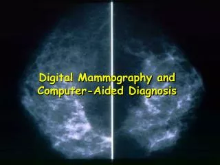
Digital Mammography and Computer-Aided Diagnosis
Breast Cancer • One out of every seven women will be diagnosed with breast cancer in 2007 • Fortunately, radical mastectomy (surgical removal) is rarely needed today with better treatment options • Breast cancer is second only to lung cancer as a cause of cancer deaths in American women
Outline • Female Breast Anatomy • Breast Cancer • Mammography • Computer-Aided Diagnosis of Breast Cancer
Muscles Muscles underneath the breasts separating them from the ribs Breast has no muscle tissue
Female Breast Anatomy • Breasts consist mainly of fatty tissue interspersed with connective tissue • There are also less conspicuous parts • lobes • ducts • lymph nodes
Breast Gland • Each breast has 15 to 20 sections (lobes) arranged like the petals of daisy • Inside each lobe are many smaller structures called lobules • At the end of each lobule are tiny sacs (bulbs) that can produce milk
Ducts 1 2 Ducts carry milk from bulbs toward dark area of skin in the center of the breast (areola) Lobes, lobules, and bulbs are Linked by a network of thin tubes (ducts) Ducts join together into larger ducts ending at the nipple, where milk is delivered Areola 3
Blood Vessels Oxygen, nutrients, and other life-sustaining nourishment are delivered to breast tissue by the blood in the arteriesand capillaries.
Lymphatic System • Lymph ducts: Drain fluid that carries white blood cells (that fight disease) from the breast tissues into lymph nodes under the armpit and behind the breastbone • Lymph nodes: Filter harmful bacteria and play a key role in fighting off infection Lymph node Lymph duct A network of vessels
Three Types of Vessels 1 Lobules Ducts Nipple Milk 3 Lymph Nodes Lymph Vessels Waste products 2 Bacteria Blood Vessels Nourishment Cell life
Signs and Symptoms Most common: lump or thickening in breast. Often painless Discharge or bleeding Redness or pitting of skin over the breast, like the skin of an orange Change in size or contours of breast Change in color or appearance of areola
Noncancerous Conditions (1) • Fibrocystic changes: Lumpiness, thickening and swelling, often associated with a woman’s period • Cysts: Fluid-filled lumps can range from very tiny to about the size of an egg • Fibroadenomas: A solid, round, rubbery lump that moves under skin when touched, occuring most in young women • Infections: The breast will likely be red, warm, tender and lumpy • Trauma: a blow to the breast or a bruise can cause a lump CBMS2006
Noncancerous Conditions (2) • Microcalcifications: Tiny deposits of calcium can appear anywhere in a breast and often show up on a mammogram • Most women have one or more areas of microcalcifications of various sizes • Majority of calcium deposits are harmless • A small percentage may be precancerous or cancer (biopsy is sometimes recommended)
Causes • Some of the cells begin growing abnormally • These cells divide more rapidly than healthy cells do and may spread through the breast, to the lymph or to other parts of the body (metastasize) • The most common type of breast cancer begins in the milk-production ducts, but cancer may also occur in the lobules or in other breast tissue CBMS2006
Normal Breast Illustration © Mary K. Bryson
Ductal Carcinoma in situ (DCIS) Carcinoma refers to any cancer that begins in the skin or other tissues that cover internal organs Ductal cancer cells Normal ductal cell Illustration © Mary K. Bryson
Invasive Ductal Carcinoma (IDC – 80% of breast cancer) • The cancer has spread to the surrounding tissues Ductal cancer cells breaking through the wall Illustration © Mary K. Bryson
Range of Ductal Carcinoma in situ Illustration © Mary K. Bryson
Invasive Lobular Carcinoma (ILC) Lobular cancer cells breaking through the wall Illustration © Mary K. Bryson
Cancer Can also Invade Lymph or Blood Vessels Cancer cells invade lymph duct Cancer cells invade blood vessel Illustration © Mary K. Bryson
Mammography • Use a low-dose x-ray system to examine breasts • Digital mammography replaces x-ray film by solid-state detectors that convert x-rays into electrical signals. These signals are used to produce images that can be displayed on a computer screen (similar to digital cameras) • Mammography can show changes in the breast up to two years before a physician can feel them
Computer-Aided Diagnosis • Mammography allows for efficient diagnosis of breast cancers at an earlier stage • Radiologists misdiagnose 10-30% of the malignant cases • Of the cases sent for surgical biopsy, only 10-20% are actually malignant CAD systems can assist radiologists to Reduce these problems National Cancer Institute
What Mammograms Show Two of the most important mammographic indicators of breat cancers • Masses • Microcalcifications: Tiny flecks of calcium – like grains of salt – in the soft tissue of the breast that can sometimes indicate an early cancer.
Detection of Malignant Masses Malignant masses have a more spiculated appearance benign malignant
Mammogram – Difficult Case • Heterogeneously dense breast • Cancer can be difficult to detect with this type of breast tissue • The fibroglandular tissue (white areas) may hide the tumor • The breasts of younger women contain more glands and ligaments resulting in dense breast tissue
Mammogram – Easier Case • With age, breast tissue becomes fattier and has fewer glands • Cancer is relatively easy to detect in this type of breast tissue
Different Views Side-to-Side MRI - Cancer can have a unique appearance – many small irregular white areas that turned out to be cancer (used for diagnosis) Top-to-Bottom
Scalar Field • A scalar field is a n-dimensional space with a scalar value attached to each point in the space (e.g., a gray-scale image)
Scalar Field and Gradient Black representing Higher values • A scalar field is a n-dimensional space with a scalar value attached to each point in the space (e.g., a gray-scale image) • The derivative of a scalar field results in a vector field called the gradient • i.e., the gradient is a vector field • which points in the direction of the greatest rate of increase of the scalar field, and • whose magnitude is the greatest rate of change
Gradient Black representing Higher values The derivative of a scalar field results in a vector field called the gradient • i.e., the gradient is a vector field • which points in the direction of the greatest rate of increase of the scalar field, and • whose magnitude is the greatest rate of change
Cartesian Gradient For an image function I(P) where P is a pixel, the Cartesian gradient at P is: P Magnitude: Orientation:
Radial Gradient Radial gradient • The radial gradient vector has the same magnitude as the Cartesian gradient vector, but • the orientation is given as: P
Feature: Spiculation [Huo et al.] • Extract the mass using a region-growing technique • The maximum gradient and its angle relative to the radial direction are computed • Calculate the full-width at half-maximum (FWHM) from the cumulative gradient orientation histogram
Feature: Spiculation [Chan et al.] • Determine the outline of the segmented mass • Obtain the rubber-band-straightening-transformed image • The spicules become approximately aligned in a similar direction • The rectangular region can then be subjected to texture analysis
Breast Calcifications • Calcifications show up as white spots on a mammogram • Round well-defined, larger calcifications (left column) are more likely benign • Tight cluster of tiny, irregularly shaped calcifications (right column) may indicate cancer
Calcification Features • The morphology of individual calcification, e.g., shape, area, and brightness • The heterogeneity of individual features characterized by the mean, the standard deviation, and the maximum value for each feature. • Cluster features such as total area, compactness
Database Approach toComputer-Aided Diagnosis • The database consists of a large number of images with verified pathology results • Diagnosis is done by submitting the suspected mass region as a query to retrieve similar cases from the database • Content-based image retrieval techniques can provide radiologists “visual aids” to increase confidence in their diagnosis
A Mammography CAD System[Giger et al.] Probability of malignancy Similar images of known diagnosis Indicates the unknown lesion relative to all lesions in the database