
Case: RUQ pain
E N D
Presentation Transcript
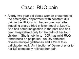
Case: RUQ pain • A forty-two year old obese woman presented to the emergency department with constant dull pain in the RUQ which began one hour after ingesting a large fried chicken meal at Luby’s. She has noted indigestion in the past and has been hospitalized only for the birth of her four children. She is febrile to 100F, has mild RUQ tenderness on palpation. An US obtained reveals multiple gallstones and a 2mm thick gallbladder wall. An injection of Demerol prior to her US completely relieved her pain.
Key to slides (left to right) • Slide 1 1U/S, acute cholecystitis, stone impacted in cystic duct 2 U/S, transverse, acute cholecystitis, thick GB wall, stones with shadowing 3 U/S, longitudinal, acute cholecystitis, thick GB wall
Key to slides (left to right) • Slide 2 1 CT scan, acute cholecystitis, thick GB wall, stones in GB 2 CT scan, acute cholecystitis, thick GB wall
Key to slides (left to right) • Slide 3 1 CT scan, emphysematous GB, air in GB wall 2 CT scan, hepatic abscess, complication of CBD obstruction 3 CT scan, intrahepatic duct obstruction from stone in CBD
Key to slides (left to right) • Slide 4 1 ERCP, gallstones (filling defects) in GB 2 IOC, choledocholithiasis
Key to slides (left to right) • Slide 5 1 PTC, intra- and extra-hepatic bile duct obstruction from distal CBD stone/tumor 2 ERCP, choledocholithiasis in distal CBD 3 ERCP, basket stone extraction
Key to slides (left to right) • Slide 6 1 Oral cholecystogram with stones 2 HIDA, nonvisualization of GB
Key to slides (left to right) • Slide 7 1 One hour HIDA scan, nonvisualization of GB
Key to slides (left to right) • Slide 8 1 Gross, GB with GS 2 Micro, acute cholecystitis, infiltration of PMNs 3 Micro, chronic cholecystitis, atrophic mucosa/thick GB wall 4 Micro, biliary obstruction, bile duct pigment