
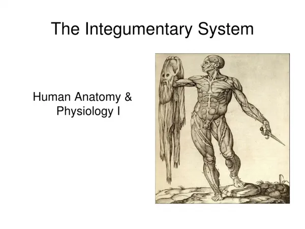
The Integumentary System
Human Anatomy & Physiology I. The Integumentary System. Introduction. Integumentary (inte: whole; gument: body covering) Helps protect the body, helps maintain a constant body temperature, and provides sensory information about the environment.

The Integumentary System
E N D
Presentation Transcript
Human Anatomy & Physiology I The Integumentary System
Introduction • Integumentary (inte: whole; gument: body covering) • Helps protect the body, helps maintain a constant body temperature, and provides sensory information about the environment. • Of all the body’s organs, none is more easily inspected or more exposed to infection, disease, and injury than the skin.
Functions of the skin • Helps regulate body temperature • Serves as water repellent • Protective barrier between the external environment and internal tissues
Functions of the skin • Excretes a small amount of salts and several organic compounds • Some capacity to absorb substances • Helps synthesize the active form of vitamin D
Two (2) ways: By liberating sweat By adjusting the flow of in the dermis Thermoregulation
Bacterial infection Mechanical injury Chemical injury Dehydration UV radiation Protection
Tactile sensations Tactile touch Pressure Vibration Tickling Thermal sensations Pain Cutaneous Sensations
Elimination of substances from the body Water evaporation (about 400 mL/daily) Sedentary person loses and additional 200 mL per day as sweat Sweat (vehicle for excretion of salts, CO2, ammonia & urea) Excretion
Certain lipid-soluble materials: Fat soluble vitamins Certain drugs Gases (O2 & CO2) Toxic materials (mercury) Toxins (poison ivy) Absorption
Requires activation of a precursor molecule in the skin (UV rays) Enzymes in liver & kidney modify the activated molecule finally producing calcitriol (most active form of vitamin D) Calcitriol is a hormone that aids in the absorption of calcium in the GI tract Synthesis of vitamin D
Epidermis (epi: above) Dermis Hypodermis (hypo: below) Structures of the skin
The epidermis is keratinized stratified squamous epithelium. 4 types of cells 4 to 5 “strata” or layers (depending on the location) Epidermis
Keratinocytes: most of the skin cells Tactile (Merkel) cells: receptor/sensation Melanocytes: production of the pigment melanin Langerhans: immune response Types of cells
Stratum basale Stratum spinosum Stratum granulosum Stratum lucidum* Stratum corneum Layers of the epidermis
Deepest layer (closest to dermis) Single row of cuboidal or columnar keratinocytes Some cells in the layer are stem cells (undergo cell division to continually produce new keratinocytes) Stratum Germinativum (Basale)
Superficial to stratum basale 8 to 10 layers of keratinocytes Projections of both Langerhans and melanocytes also appear in this stratum Stratum spinosum
3 to 5 layers of flat keratinocytes Marks the transition between the deeper, metabolically active strata and the dead cells of the more superficial strata. Stratum granulosum
Present only in the thick skin of the fingertips, palms, and soles 3 to 5 layers of flattened clear, dead keratinocytes Large amount of keratin Stratum lucidum*
25 to 30 layers of flattened dead keratinocytes. Cells are continuously shed and replaced by cells from the deepest strata. Constant exposure of skin to friction stimulates the formation of a callus (abnormal thickening of the stratum corneum). Stratum corneum
Composed mainly of connective tissue containing and elastic collagen fibers. Blood vessels, nerves, glands, and hair follicle are embedded in the in dermal tissue. Dermis
Two layers: Papillary layer Reticular layer Dermal papillae: extensions of the dermis into the epidermis forming the ridges of the fingerprints Dermis
Deep to the dermis, but not part of the skin! Mostly adipose tissue Serves as a storage depot for fat and contains large blood vessels that supply the skin. Hypodermis
Hair on the head guards the scalp from injury and the sun’s rays. Decreases heat loss from the scalp. Eyelashes and eyebrows protect the eyes from foreign particles. Sensory (touch receptors) Hair
Nails are plates of tightly packed, hard, keratinized epidermal cells. Nail body: visible part Free edge: extend past the distal end of the digit Nail root: portion that is buried in a fold of skin Lunula: whitish, crescent-shaped Nails
There are two main types: eccrine and apocrine sweat glands. The cells of sweat glands release their secretions by exocitosis and empty them into hair follicles of onto the skin surface through pores. Sweat (sudoriferous) gland
Eccrine sweat glands • Distribution: Throughout the skin • Location of secretory portion: mostly deep in dermis • Termination of excretory duct: surface of epidermis • Secretion: less viscous; consist of water, ions, urea, uric acid, ammonia, glucose, and lactic acid • Functions: thermoregulation, and waste removal • Onset of function: soon after birth
Apocrine sweat glands • Distribution: skin of axilla, groin, areola, and bearded regions of face • Location of secretory portion: mostly in subcutaneous layer • Termination of excretory duct: hair, follicle • Secretion: more viscous; consist of same components as eccrine sweat glands plus lipids and proteins • Functions: stimulated during emotional stress and sexual excitement • Onset of function: puberty
Usually connected to hair follicles. They are absent in the palms and soles. Sebaceous glands produce sebum, which moistens hairs and waterproofs the skin. Clogged sebaceous glands may produce acne. Sebaceous (oil) glands
Hyperthermia • Increased blood and internal temperature. • Increased temperature is sensed by the hypothalamus. • Vasodilation occurs in skin blood vessels so more heat is lost from the skin. • Sweat glands become active, increasing evaporative heat loss. • Body temperature decreases!
Hypothermia • Decreased blood and/or skin temperature. • Decreased temperature is sensed by the hypothalamus. • Vasoconstriction occurs in skin blood vessels so less heat is loss to the environment. • Skeletal muscles are activated, causing shivering, which increases metabolism and generates heat. • Body temperature increases.


