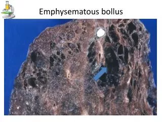
Emphysematous cystitis
Emphysematous cystitis. James Montgomery, DVM April 27, 2009. Marta. 12 years old Female Bichon frise Hx: recurrent UTIs, hematuria, stranguria, renoliths, struvite uroliths, hyperadrenocorticism, diabetes mellitus. Radiographs: Acc# 78616. Gas opacities superimposed with bladder

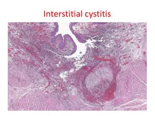
Emphysematous cystitis
E N D
Presentation Transcript
Emphysematous cystitis James Montgomery, DVM April 27, 2009
Marta • 12 years old • Female • Bichon frise • Hx: recurrent UTIs, hematuria, stranguria, renoliths, struvite uroliths, hyperadrenocorticism, diabetes mellitus
Radiographs: Acc# 78616 • Gas opacities superimposed with bladder • Marked hepatomegaly • Soft tissue mineralization • Bilateral stifle djd
Ultrasound: Acc# 78607 • Gas shadowing from within urinary bladder walls – not gravity dependent • Mucosal irregularities or calculi • Bilateral peripelvic mineralization • Possible splenic mineralization • Hypoechoic hepatomegaly
Emphysematous cystitis • In humans, reported at a higher incidence in females • Usually diabetic patients • Three radiographic stages • 1: A clear 1 mm zone seen around the contrast medium and free gas not present in the bladder lumen • 2: Bladder wall is irregular and thickened because of increased intramural gas. Still no free intraluminal gas. • 3: Free gas in the bladder lumen evident radiographically.
Emphysematous cystitis • First reported in a diabetic dog in 1926 • Reported with approx. same frequency in males and females • Occurs mainly in association with diabetes, but also with primary renal glucosuria. • Glucose-fermenting bacteria or yeast ferment glucose to produce carbon dioxide small gas bubbles which collect in the bladder wall and lumen. • Gas bubbles coalesce and rupture as the cystitis progresses
Emphysematous cystitis • In nonglucosuric dogs: • Develops secondary to other conditions – • Chronic UTI • Bladder trigone diverticulum • Chronic steroid administration • Production of gas is due to bacterial breakdown of urinary albumin by bacteria • Bladder wall hypoxia • Common bacteria: Proteus mirabilis (all 4 dogs in Petite article), Escherichia coli, Aerobacteraerogenes, Clostridium sp. (more specifically perfringens)
Emphysematous cystitis • Treatment effective treatment of UTI and control of glucosuria
References • Adams LG, Syme HM. Canine Lower Urinary Tract Diseases. In Ettinger SJ, Feldman EC, eds. Textbook of Veterinary Internal Medicine, 6thed (St. Louis, MO: Elsevier Saunders, 2005) p. 1872. • Nyland TG, et al. Urinary Tract. In Nyland TG, Mattoon JS, eds. Small Animal Diagnostic Ultrasound, 2nded (Philadelphia, PA: Saunders Elsevier, 2002) p. 183. • Petite A, et al. Radiographic and ultrasonagraphic findings of emphysematous cystitis in four nondiabetic female dogs. Veterinary Radiology & Ultrasound 2006;47(1):90-3.