Download
1 / 15
150 likes | 410 Vues
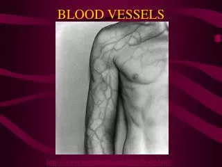
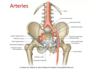
7.4 – Blood Vessels Arteries arteries carry blood away from the heart they have thick walls with distinct layers outer and inner connective tissue middle muscle and elastic connective tissue

E N D
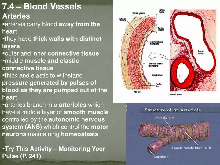
7.4 – Blood Vessels • Arteries • arteries carry blood away from the heart • they have thick walls with distinct layers • outer and inner connective tissue • middle muscle and elastic connective tissue • thick and elastic to withstand pressure generated by pulses of blood as they are pumped out of the heart • arteries branch into arterioles which have a middle layer of smooth muscle controlled by the autonomic nervous system (ANS) which control the motor neurons maintaining homeostasis • Try This Activity – Monitoring Your Pulse (P. 241)
Capillaries • composed of a single layer of cells, capillaries are the site of fluid and gas exchange between blood and body cells • most are 0.4-1.0 mm long and less than 0.005 mm in diameter (RBC must pass through in single file)(Fig. 3, P. 252) • capillary beds are easily damaged by high BP or impact (bruising occurs as blood enters space between tissue) • O2 and CO2 diffuse across capillary walls • proteins cross by active transport (exocytosis and endocytosis) • water-soluble ions and vitamins pass through spaces between endothelial cells
contraction of smooth muscle causes vasoconstriction, decreasing blood flow to tissues • relaxation of smooth muscles causes vasodilation, increasing the flow of blood to tissues and allowing for local cells to perform energy-consuming tasks • precapillary sphincter muscles regulate the movement of blood from arterioles into capillaries • relax to allow blood flow only when needed (1 of 50 capillaries open at any time)
7.10 – Capillary Fluid Exchange • between the blood and ECF • O2 and CO2 diffuse across capillary walls • proteins cross by active transport (exocytosis and endocytosis) • water-soluble ions and vitamins pass through spaces between endothelial cells
interstitial fluid pressure and capillary osmotic pressure regulate the movement of water between blood and ECF • water and small mineral ions move from the higher pressure of capillaries into the much lower pressure of ECF (filtration) • proteins, RBC and WBC remain in the capillary • osmotic pressure moves fluid back into capillaries as higher solute (protein) concentration creates a hypertonic condition relative to ECF (absorption)
the balance between osmotic pressure and fluid pressure is upset during hemorrhage, starvation, or inflammation • decrease in blood volume during hemorrhage lowers BP • pressure driving fluid from capillaries into ECF decreases, but osmotic pressure in ECF remains the same • net movement of fluid into capillaries provides a homeostatic adjustment
starvation causes the use of plasma proteins as a last resort • decreased number of proteins lowers osmotic pressure, decreasing absorption • more water enters tissue space than is pulled back into capillaries, causing edema (swelling) • inflammation or allergic reactions cause the release of histamine • capillary permeability increases, allowing antibodies and WBC to search for “pathogen” • change in osmotic pressure opposes the one in capillaries, reducing absorption and resulting in swelling • anaphylactic reactions are severe allergic reactions that present very quickly and can be expedited by adrenaline or antihistamines
Veins • deoxygenated blood collects in small veins (venules) as capillaries merge
BP in venules is reduced to 15-20 mm Hg • thinner walls of veins contain smooth muscle that rythmically massage blood back to the heart
valves in veins prevent backflow (act like a one-way door) • sketetal muscle also aids venous blood flow as contraction decreases vein diameter and pressure increases (Fig. 4, P. 253) • sequential contractions massage blood back to heart (stretching in the morning stimulates blood flow, long periods of standing motionless results in faintness) • under stress, smooth muscle contractions can move more blood to the heart to increase energy supply • pooling of blood in veins can result in the one-way valves failing • gravity carries blood toward feet where more pooling occurs varicose veins
Identification of Venous Valves Role up your sleeve so you can see the veins on the palmar side of the forearm. Place your forefinger on one of the veins evident, then push the thumb along the vein toward the shoulder. Leave the finger in place, and observe if the blood flows back into the vein. Now remove the finger, and observe what happens. Place the finger on one of the veins (preferably the same vein) and push the thumb along the vein toward the hand. Leave the finger in place, and observe if the blood flows back into the vein. Now remove the finger, and observe what happens. Based on these observations, predict where the valves are in the vein. You may choose to repeat this on of the veins on the top of the hand.
7.8 – Blood Pressure • the force of blood on the walls of the arteries • can be measured by a sphygmomanometer • air bladder is inflated to cut off flow to the brachial artery • air is slowly released until a pulse is heard (stethoscope) and seen (pressure gauge) as blood pressure overcomes air pressure • air is continued to be released until pulse cannot be heard or seen • systolic pressure measures pressure during ventricular contraction • diastolic pressure measures pressure during ventricular relaxing • N.B. “normal” systolic/diastolic BP is 120/80 measured in mm Hg
BP is determined by cardiac output and arterial resistance • smooth muscle around arteries regulates blood flow (and therefore pressure) in response to neural and hormonal controls, as well as metabolic products • i.e. when BP exceeds acceptable levels, receptors in the aorta and carotid arteries send a signal to the medulla oblongata in the brain • sympathetic nerve impulses dilate arteries, parasympathetic nerve impulses decreases heart rate and stroke leading to decreased blood pressure • accumulation of CO2and lactic acid (due to cellular respiration) causes smooth muscles to relax • vasodilation increases blood flow to tissues, delivering more oxygen
7.9 – Variation in Cardiovascular Output • cardiac disease includes hypertension (sustained high blood pressure), arteriosclerosis, atherosclerosis, heart attack (destruction of the heart muscle), and stroke (interrupted blood flow to the brain) • factors affecting health of the heart: • high blood cholesterol • smoking • diabetes mellitus • hypertension • sedentary lifestyle • rapid weight gain or loss • congenital factors • Hypertension • increased resistance to blood flow results in weakening and rupture of blood vessels • increased connective tissue to support weak vessels makes them less elastic which further increases blood pressure • sometimes hereditary , but usually diet-related (i.e. salt )
7.11 – The Lymphatic System • lymph is a plasma-like fluid that drains leaked proteins from the ECF in vein-like vessels to the venous system • lymph nodes at intervals filter damaged cells and debris, house WBC that engulf bacteria, and supply lymphocytes • the spleen filters lymph and is richly supplied with blood sinuses, where specialized blood cells enter the circulatory system • the thymus gland is where T lymphocytes mature • as a fetus or early postnatal, proteins enter thymus and act as antigens activating T cells • once activated, T cells are destroyed, ensuring that remaining thymic cells cannot produce antibodies against body proteins