Download
1 / 83
840 likes | 1.01k Vues
Explore the intricate structure, tracts, and syndromes of the spinal cord, including the roles of meninges, vascular supply, and internal organization. Gain insights into the functional divisions and cellular composition of the gray and white matter in the spinal cord.

E N D
The Spinal Cord • Structure of the spinal cord • Tracts of the spinal cord • Spinal cord syndromes Nabeel Kouka, MD, MBA, FABMISS www.brain101.info
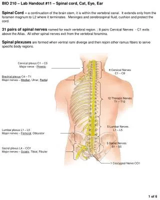
Spinal Cord - Comparable to Input-Output (IO) System of the Computer - Spinal Nerve (C8, T12, L5, S5, Cx1) - Segmental Structure of Neural Tube Origin
Spinal segment C8, T12, L5, S5, Cx1 Anterior (Ventral) Root Posterior (Dorsal) Root Dorsal Root (Spinal) Ganglion Root - Rootlets
Conus Medullaris (L1-2) Cauda Equina Anterior median fissure Anterolateral fissure
Posterior median sulcus Posterolateral sulcus Posterior intermediate sulcus Fasciculus cuneatus Fasciculus gracilis Posterior surface of the spinal cord
Spinal Cord Meninges Periosteum of Vertebra - Epidural Space ----------------- epidural anesthesia Dura Mater Spinalis Arachnoid Membrane - Subarachnoid Space -------- Lumbar Puncture Spinal Anesthesia Pia Mater Spinalis - Denticulate Ligament --------- Cordotomy - Filum Terminale
Meninges of • the spinal cord • Dura mater • Arachnoid membrane • Pia mater Denticulate ligament - specialization of the pia mater - landmark for cordotomy
Spinal Cord Vascular Supply Arterial Supply - Spinal Arteries Anterior (1) & Posterior (2) Spinal Artery from Vertebral artery - Radicular Arteries ----- Segmental arteries from Vertebral, Ascending Cervical, Intercostal and Lumbar Artery Venous Drainage - Longitudinal & Radicular Veins to Intervertebral veins ---- to Internal Vertebral Venous Plexus to external vertebral venous plexus ---- to segmental veins
5. Adamkiwicz artery anterior spinal artery segmental arteries
Spinal Cord External Figure Conus Medullaris (L1-2) Spinomedullary Junction - Foramen Magnum, Pyramidal decussation, C1 ventral root Enlargements - Cervical (C4-T1) & Lumbosacral (L1-L4) Longitudinal Fissures - anterior median fissure - anterolateral fissure - posterior median sulcus - posterolateral sulcus
cervical enlargement (C8) thoracic cord (T8) lumbar enlargement (L3) sacral cord (S1)
Cervical level • Wide flat cord, lots of white matter, • ventral horn enlargements. • Lumbar level • - Round cord, ventral horn enlargements. • Sacral level • - Small round cord, lateral Horn. • Tthoracic level • - Notice the pointed tips which stick out • between the small dorsal and ventral horns. • This extra cell column is called the • intermediate horn (AKA: Intermediolateral • Cell Column). It is the source of all of the • sympathetics in the body & occurs only in • the Thoracic sections T 1 - L 2
Spinal Cord Internal Structure White Matter Anterior Funiculus (Anterior White Column) Posterior Funiculus (Posterior White Column) Fasciculus Gracilis & Fasciculus Cuneatus Lateral Funiculus (Lateral White Column) Gray Matter Anterior Horn ------------ --- motor Posterior Horn -------------- sensory Lateral Horn ----------------- autonomic (sympathetic) Gray Commissure -------- anterior and posterior
1. posterior horn 2. anterior horn 3. intermediate zone (intermediate gray) 4. lateral horn 5. posterior funiculus 6. anterior funiculus 7. lateral funiculus 8. Lissauer's tract 9. anterior median fissure 10. posterior median sulcus 11. anterolateral sulcus 12. posterolateral sulcus 13. Posterior intermediate sulcus
Spinal Cord Internal Structure Principles of Cord Organization 1) Longitudinal Arrangement Fibers (White Matter) ------------- White Column Cell Groups (Gray Matter) ------- Gray Column 2) Transverse Arrangement Afferent & Efferent Fibers Crossing (Commissural and Decussating) Fibers 3) Somatotopical Arrangement
Lamina of Rexed Lamina I Posteromarginal Nucleus Lamina II Substantia Gelatinosa of Rolando Lamina III Lamina IV, V, VI ----- Nucleus Proprius Lamina VII - Intermediate Gray - Intermediolateral cell column (ILM) - Clarke’s column (Nucleus dorsalis) - Intermediomedial cell column (IMM) Lamina VIII Lamina IX ---------- Anterior Horn (Motor) Cell Lamina X ----------- Gray Commissure
Lamina I • AKA: lamina marginalis • or the layer of Waldeyer • Receives incoming dorsal root fibers • and collateral branches as well • Larger neurons contribute axons • to Contralateral Spinothalamic Tract
Lamina II • AKA: Substantia Gelatinosa • Involved in Pain interpretation • Receives incoming input from dorsal • rootaxons & descending input from • reticular formation of the medulla • Efferent axons travel up & down • several segments to make contact • with other areas of the dorsal horn
Lamina III • Contains larger, less densely packed • cells than lamina II • Receives primary afferents from • dorsal root fibers • Neurons considered as a part of • nucleus proprius
Lamina IV • Contains a variety of cell types that have • more myelin than any other lamina • Some tract cells originate here, axons cross • the midline and enter the contralateral • Spinothalamic Tract, also sends contacts to • layers II and III • Receives afferents from dorsal roots via • the dorsal funiculus • At rostral end of spinal cord, laminas I-IV • become continuous with the spinal • trigeminal nucleus
Lamina V - VI • Origination of tract cells, similar • to lamina IV, these tracts cells are • also known as the Nucleus Proprius • (e.g. spinal thalamic tract or • anterolateral system; pain and • temperature, some tactile) • Receives afferent input from • dorsal roots and descending fibers, • most importantly Corticospinal
Lamina V - VI C7 reticular formation----------------> Laterally, gray matter at base of dorsal horn mixes with white matter from lateral funiculus, this region is called reticular formation. It is noticeable in the cervical region
Lamina VII • The largest region, occupies most of • ventral horn &intermediate zone • Projects long axons that connect to other • gray matter segments of the cord • Some columns do not fit into the lamina • scheme, and have individual designations: • a. Nucleus dorsalis (Clarke) • b. Intermediolateral cell column • c. Intermediomedial cell column • d. Sacral autonomic nucleus
Lamina VII • Nucleus dorsalisof Clark • AKAnucleus thoracicus • is located medial & ventral • to the dorsal horn in T1-L3 • Composed of large neurons • & axons that form the • dorsal spinocerebellar tract • on the ipsilateral side T5
Lamina VII • Intermediolateral cell column • is located at the lateral portion • of the intermediate zone. • Responsible for the formation • of the lateral horn in T1 - L2 • Consists of cell bodies of • sympathetic preganglionic • neurons T5
Lamina VII • Intermediomedial cell column • is located lateral to lamina X. • Not seen in all cord sections. • Receives primary afferent • fibers from dorsal root and • has been implicated in • visceral reflexes T5
Lamina VII S2 • Sacral autonomic nucleus • is located in the lateral part of • lamina VII in S2-S4 segments • Consists of preganglionic para- • sympathetic neurons
Lamina VIII • Located on the medial aspect of • the ventral horn • Efferent projections both ipsilaterally • and contralaterally to the same and • nearby segmental levels to lamina • VII & IX • Site of termination for descending • fibers, including the vestibulospinal • and reticulospinal tracts
Lamina IX • Consists of columns of neurons • embedded in either lamina VII or VIII • Cells include alpha and gamma motor • neurons, which axons exit via the • ventral roots and innervate striated • muscle. Smaller neurons contribute • axons to the ventral fasciculus proprius • Four columns of motor neurons can • be identified within this lamina; • ventromedial, ventrolateral, dorsolateral • & central each has characteristic • dendritic features
Lamina IX Ventral gray columns in lamina IX have somatotopic arrangement: - Medial areas innervate the axial musculature - Lateral areas innervate the limbs muscles
PHRENIC NUCLEUS The phrenic nucleus is located in the ventromedial area of the ventral horn in C2-C5 segments. It receives bilateral innervation from the solitary nucleus of the medullary region, via solitary tract. This nucleus is responsible for the innervation of the diaphragm SPINAL ACCESSORY NUCLEUS The spinal accessory nucleus (cranial nerve XI) is located in the lateral area of vental horn in C1-C5 segments. Corticospinal tract innervates this nucleus bilaterally. This nucleus is also responsible for the innervation of the trapezius & sternocleidomastoid muscles (ipsilaterally)
Lamina IX • Located ventrolaterally in • S1-S2 spinal segments • Supplies muscles of the • pelvic floor, including striated • muscle sphincters for urinary • and fecal continence Nucleus of Onuf S2
Lamina X • Surrounds the central canal, and • includes the ventral gray commissure • Contains relatively small neurons, • radial neuroglia cells & decussating • axons • Some dorsal root afferents terminate • here
Fasiculus Proprius • Ascending and descending association fiber systems of the • spinal cord which lie deep in the anterior, lateral & posterior • funiculi adjacent to the gray matter. • Fasciculi proprii aka Flechsig's fasciculi or Ground bundles • consist of: anterior, lateralis & intersegmental fasciculi
Dorsal Roots • Each dorsal root divides • into 6 - 8 rootlets • Each rootlet can be divided into • lateral & medial division • Lateral division carries • information related to pain • & temperature • Medial division carries • information related to tactile • discrimination & vibration
Dorsal Roots • Lateral divisionaxons enter • dorsolateral tract of Lissauer, • and then divide into ascending • & descending branches, each • terminate in the dorsal horn • Most terminate at same • level & some fibers may • travel up or down the cord • up to four levels
Dorsal Roots • Medial division axons enter the • white matter & then divide into • ascending & descending branches • Descending branches are • organized into two bundles, • the Septomarginalis Fasiculus • and the Interfascicular Fasiculus • All descending branchesterminate • in the dorsal horn
Dorsal Roots • Ascending branches of the • medial divisionenter the • dorsal funiculus & terminate • in gracile & cuneate nuclei • in the medulla
Ventral Horn • Lamina IX contains two types • of motor neurons, alpha and • gamma • Alpha motor neurons innervate • extrafusal fibers of striated • skeletal muscles • Gamma motor neurons innervate • intrafusal fibers of neuromuscular • spindles • Both types receive inputs from • interneurons, including the • inhibitory Renshaw cell
Tracts of the Spinal Cord Fasciculus Gracilis Fasciculus Cuneatus Tractus spinocerebellaris dorsalis Tractus corticospinalis lateralis Tractus spinothalamicus lateralis Tractus spinocerebellaris ventralis Tractus rubrospinalis Tractus spinotectalis Tractus corticospinalis anterior Tractus olivospinalis Tractus spinoolivaris Tractus tectospinalis Tractus reticulospinalis Tractus vestibulospinalis Tractus spinothalamicus anterior Raphe-spinal & Hypothalamospinal fibers 16
Spinal Cord Tracts Ascending Tracts Modality: Touch, Pain, Temperature, Kinesthesia Receptor: Exteroceptor, Interoceptor, Proprioceptor Primary Neuron: Dorsal Root Ganglion (Spinal Ganglion) Secondary Neuron: Spinal Cord or Brain Stem Tertiary Neuron: Thalamus (Ventrobasal Nuclear Complex) Termination: Cerebral Cortex, Cerebellar Cortex, or Brain Stem
Spinal Cord Tracts Ascending Tracts Posterior White Column-Medial Lemniscal Pathway Spinothalamic Tract Spinoreticular or Spinoreticulothalamic Tract Spinocerebellar Tract Spinomedullothalamic Tract Cervicothalamic or Spinocervicothalamic Tract Spino-olivary Tract Spinotectal Tract