Download
1 / 6
60 likes | 290 Vues
BIO 210 – Lab Handout #11 – Spinal cord, Cat, Eye, Ear Spinal Cord – a continuation of the brain stem, it is within the vertebral canal. It extends only from the foramen magnum to L2 where it terminates. Meninges and cerebrospinal fluid, cushion and protect the cord.

E N D
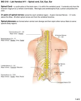
BIO 210 – Lab Handout #11 – Spinal cord, Cat, Eye, Ear Spinal Cord – a continuation of the brain stem, it is within the vertebral canal. It extends only from the foramen magnum to L2 where it terminates. Meninges and cerebrospinal fluid, cushion and protect the cord. 31 pairs of spinal nerves named for each vertebral region. ; 8 pairs Cervical Nerves - C1 exits above the Atlas. All other spinal nerves exit from the vertebral foramina. Spinal plexuses are formed when ventral rami diverge and then rejoin other ramus fibers to serve specific body regions. Cervical plexus C1 – C5 Major nerve - Phrenic 8 Cervical Nerves C1 – C8 Brachial plexus C4 – T1 Major nerves – Median, Ulnar, Radial 12 Thoracic Nerves T1 – T12 Lumbar plexus L1 – L5 Major nerves – Femoral, Obturator 5 Lumbar Nerves L1 – L5 5 Sacral Nerves S1 – S5 Sacral plexus L4 – CO1 Major nerves – Sciatic, Tibial, Fibular 1 Coccygeal Nerve CO1 1 of 6
Nerves to identify on Model & Cat Brachial plexus Radial nerve Vagus nerve http://biology.clc.uc.edu/fankhauser/Labs/Anatomy_&_Physiology/A&P202/Nervous_System_Anatomy/cat_nerve_jpegs/brachial_plexus_P1281871.JPG http://biology.clc.uc.edu/fankhauser/Labs/Anatomy_&_Physiology/A&P202/Nervous_System_Anatomy/cat_nerve_jpegs/radial_nerve_P1281874.JPG http://biology.clc.uc.edu/fankhauser/Labs/Anatomy_&_Physiology/A&P202/Nervous_System_Anatomy/Cat_Nerve_jpegs/vagus_nerves_P1281875.JPG Sciatic nerve Phrenic nerve http://biology.clc.uc.edu/fankhauser/Labs/Anatomy_&_Physiology/A&P202/Nervous_System_Anatomy/Cat_Nerve_jpegs/phrenic_nerve_right_P1281879.JPG http://biology.clc.uc.edu/fankhauser/Labs/Anatomy_&_Physiology/A&P202/Nervous_System_Anatomy/Cat_Nerve_jpegs/sciatic_nerve_P1281882.JPG Brachial plexus – formed from spinal nerves C4 – T1 Radial nerve – largest nerve to exit brachial plexus; moves deep to biceps brachii Vagus Nerve, Cranial X – the only cranial to leave the head and neck. It innervates the heart, lungs, liver, gallbladder, stomach, pancreas, large and small intestines Phrenic Nerve – exits the Cervical Plexus and innervates the Diaphragm Sciatic nerve – largest nerve to exit sacral plexus; posterior to the femur 2 of 6
Gray Matter - Gray matter and white matter have reversed positions from that found in the brain. Gray matter looks like a butterfly or the letter H • Anterior (ventral) horn – contains cell bodies of motor neurons of the SNS & ANS • Posterior (dorsal) horn – contains Interneuron cell bodies for sensory nuclei. Interneurons integrate incoming sensory signals from the PNS. Does NOT reach the edge of the cord • Lateral horn – in the thoracic and lumbar regions; contains cell bodies of visceral motor neurons • Gray commissure – central bridge of gray matter connecting the two sides of the cord • Central canal – in the gray commissure; contains cerebrospinal fluid • Dorsal root – sensory axons that enter the cord from the dorsal root ganglion • Dorsal root ganglia – enlarged area containing cell bodies of sensory neurons • Ventral root – axons of the motor neurons found in the anterior (ventral) horn • Spinal nerves – formed from the fusion of the dorsal and ventral roots just beyond the dorsal root ganglion; 1-2 cm long Dorsal root ganglion Dorsal root Lateral funiculus Ventral root Spinal nerve Posterior horn Central canal Posterior median sulcus Posterior funiculus Sensory neuron axon Lateral horn Anterior horn Gray commissure Anterior funiculus Motor neuron cell body Sensory neuron cell body Anterior median fissure Motor neuron axon • White Matter - myelinated fibers divided into 3 primary regions or white columns; • 1. Posterior funiculi 2. Lateral funiculi 3. Anterior funiculi • Divided into right and left halves by 2 deep grooves: • Anterior median fissure – between the anterior horns of gray matter • Posterior median sulcus – between the posterior horns Spinal meninges Pia mater Arachnoid Dura mater 3 of 6
Special Senses: Anatomy of the Eye Extrinsic muscles – 6 attached to the exterior of the eyeball Conjunctiva – mucous membrane lining the internal surface of the eyelids and continues over the anterior surface of the eyeball the cornea Anterior cavity (Aqueous Humor) Cornea Conjunctiva Pupil Iris Ciliary body Lens Posterior cavity (Vitreous Body) Retina Choroid Sclera Extrinsic muscle Superior oblique Superior rectus Medial rectus not visible Lateral rectus The wall of the eye is constructed of three layers or tunics. 1. The outermost fibrous tunic is a protective layer of dense CT. Sclera – opaque white of the eye; Cornea – anterior region; transparent to allow the entry of light Inferior rectus Inferior oblique • The middle vascular tunic(uvea) contains 4 parts: • Choroid – posterior-most part; dark pigment prevents light scattering; richly vascular layer • Ciliary body – anterior portion of Choroid; modified for lens attachment; produces aqueous humor; changes shape of lens • Lens – flexible crystalline structure in the middle of the ciliary body • Iris – pigmented; circular and radial smooth muscle fibers open & close a diaphragm controlling the amount of light entering the eye • Pupil – rounded opening in the iris through which light passes Retina – the innermost sensory tunic; delicate two cell types: 1. Pigmented epithelial layer – nourish and support photoreceptor cells 2. Photo receptor cells – Rods and Cones. Optic disc – blind spot; site where Optic nerve leaves the eyeball The lens divides the eye into 2 compartments: Anterior cavity – lens to cornea contains Aqueous humor – clear watery fluid Posterior Cavity – behind the lens contains Vitreous humor (body) – gel-like substance 4 of 6
Anatomy of the Ear Outer or External Ear – 3 parts 1. Pinna (auricle) – skin-covered cartilaginous structure; collects and directs sound waves 2. External auditory meatus (canal) – a short, narrow chamber in the temporal bone; 3. Tympanic membrane – eardrum; membrane found at the end of the meatus; sound waves strike this membrane causing it to vibrate; Separates outer & inner ear Vestibular branch Semicircular canals Cochlear branch Oval window into Cochlea Organ of Corti within Cochlea Round window from Cochlea Semicircular canals Middle Ear – 4 parts Cavity containing ossicles that transmit the vibration of the eardrum to the fluids of the inner ear 1. Malleus (hammer) – first ossicle touching the eardrum 2. Incus (anvil) – the middle ossicle 3. Stapes (stirrup) – the innermost ossicle touching the oval window of the inner ear 4. Auditory tube – connects the middle ear chamber with the nasopharynx; equalizes the pressure of the middle ear cavity with the external air pressure Incus Vestibulocochlear nerve Pinna Malleus External acoustic meatus Cochlea Tympanic membrane Stapes Auditory tube Vestibule • Inner Ear – 3 divisions in a system of bony, tortuous chambers called the bony labyrinth which is filled with an aqueous fluid the Perilymph. Within the bony labyrinth is a membranouslabyrinth that follows the contours of the bony chambers. It is filled with a more viscous fluid called Endolymph. • 1. Semicircular Canals – the most superior division; • 2. Vestibule – middle region; • a. Oval window – point where stapes is seated; Transmits vibrations to the perilymph in the upper chamber of the Cochlea • b. Round window – where the lower chamber of the Cochlea meets the Vestibule 3. Cochlea – snail-like; a. Organ of Corti – inside the cochlear duct; contains the receptors for hearing; Stapes fits into 8 Steps to Hearing Sound waves enter the external acoustic meatus Tympanic membrane vibrates slow for deep & fast for high pitched sounds Vibrations are amplified by malleus, incus and then stapes Oval window vibrates 20X greater than tympanic membrane Fluid pressure waves are created in perilymph of scala vestibuli Fluid pressure waves continue on to perilymph of scala tympani Vibration of the vestibular membrane creates pressure waves in the endolymph and vibration of basilar membrane Hair cells are moved against the tectoral membrane bending the stereocilia and generating a nerve impulse 5 of 6
Dissection of the Cow or Sheep Eye • Put on safety glasses and gloves; Cut open plastic bag containing preserved eye and rinse with tap water; place in dissecting pan • Examine the external surface of the eye; note thick cushion of adipose tissue. Identify • Optic nerve (cranial nerve II) as it leaves the eyeball • Extrinsic eye muscles • Conjunctiva • Sclera • Cornea (normally transparent it is opaque in the preserved eye) • Trim away most of the fat and connective tissue but leave the optic nerve intact. • Hold the eye with the cornea facing downward • About ¼ inch above the cornea, make an incision with a sharp scalpel into the sclera • Use scissors to complete the incision around the circumference of the eyeball parallel to the corneal edge • Carefully lift the anterior part of the eyeball away from the posterior portion. The vitreous body should remain with the posterior part of the eyeball • Examine the anterior part of the eye and identify the following: • Ciliary body – black pigmented body that appears to be a halo encircling the lens • Lens – biconvex structure that is opaque • Suspensory ligament – a halo of delicate fibers attaching the lens to the ciliary body • Carefully remove the lens, note the aqueous humor and identify the adjacent structures • Iris – anterior continuation of the ciliary body penetrated by the pupil • Cornea – more convex anterior most portion of the sclera; normally transparent but cloudy in preserved specimens • Examine the posterior portion of the eyeball. Remove the vitreous humor and identify the following: • Retina – neural layer of the retina appears as a delicate white, probably crumpled membrane that separates easily from the pigmented choroid • Optic disc – where the retina is attached and the optic nerve leaves the eyeball • Choroid – pigmented coat appears iridescent in the cow or sheep eye due to a special reflecting surface call the tapetum lucidum. It is not found in humans 6 of 6