The Peripheral Nervous System (PNS)
1.18k likes | 2.05k Vues
The Peripheral Nervous System (PNS). P A R T A. Peripheral Nervous System (PNS). PNS – all neural structures outside the brain and spinal cord Includes sensory receptors, peripheral nerves, associated ganglia, and motor endings Provides links to and from the external environment.

The Peripheral Nervous System (PNS)
E N D
Presentation Transcript
The Peripheral Nervous System (PNS) P A R T A
Peripheral Nervous System (PNS) • PNS – all neural structures outside the brain and spinal cord • Includes sensory receptors, peripheral nerves, associated ganglia, and motor endings • Provides links to and from the external environment
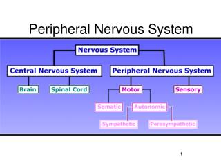
PNS in the Nervous System Figure 13.1
Sensory Receptors • Structures specialized to respond to stimuli • Activation of sensory receptors results in depolarizations that trigger impulses to the CNS • The realization of these stimuli, sensation and perception, occur in the brain
Receptor Classification by Stimulus Type • Mechanoreceptors – respond to touch, pressure, vibration, stretch, and itch • Thermoreceptors – sensitive to changes in temperature • Photoreceptors – respond to light energy (e.g., retina) • Chemoreceptors – respond to chemicals (e.g., smell, taste, changes in blood chemistry) • Nociceptors – sensitive to pain-causing stimuli
Receptor Class by Location: Exteroceptors • Respond to stimuli arising outside the body • Found near the body surface • Sensitive to touch, pressure, pain, and temperature • Include the special sense organs
Receptor Class by Location: Interoceptors • Respond to stimuli arising within the body • Found in internal viscera and blood vessels • Sensitive to chemical changes, stretch, and temperature changes
Receptor Class by Location: Proprioceptors • Respond to degree of stretch of the organs they occupy • Found in skeletal muscles, tendons, joints, ligaments, and connective tissue coverings of bones and muscles • Constantly “advise” the brain of one’s movements
Receptor Classification by Structural Complexity • Receptors are structurally classified as either simple or complex • Most receptors are simple and include encapsulated and unencapsulated varieties • Complex receptors are special sense organs
Simple Receptors: Unencapsulated • Free dendritic nerve endings • Respond chiefly to temperature and pain • Merkel (tactile) discs • Hair follicle receptors
Simple Receptors: Encapsulated • Meissner’s corpuscles (tactile corpuscles) • Pacinian corpuscles (lamellated corpuscles) • Muscle spindles, Golgi tendon organs, and Ruffini’s corpuscles • Joint kinesthetic receptors
Unencapsulated Receptors Table 13.1.1
Simple Receptors:Encapsulated Table 13.1.2
From Sensation to Perception • Sensation is the awareness of changes in the internal and external environment • Perception is the conscious interpretation of those stimuli
Organization of the Somatosensory System • Input comes from exteroceptors, proprioceptors, and interoceptors • The three main levels of neural integration in the somatosensory system are: • Receptor level – the sensor receptors • Circuit level – ascending pathways • Perceptual level – neuronal circuits in the cerebral cortex
Processing at the Receptor Lever • The receptor must have specificity for the stimulus energy • The receptor’s receptive field must be stimulated • Transduction • Conversion of the energy of a stimulus into the energy of a nerve signal
Processing at the Receptor Lever • Receptor potential • It is a graded potential happening on a receptor • Depolarization or hyperpolarization • Generator potential • It is a receptor potential strong enough to cause an action potential in an afferent fiber
Adaptation of Sensory Receptors • Adaptation is a reduction in sensitivity in the presence of a stimulus • Receptor membranes become less responsive • Receptor potentials decline in frequency or stop
Adaptation of Sensory Receptors • Tonic receptors • Have little peripheral adaptation • Chemical interoceptors • Pain receptors • Macula in the vestibular apparatus • Proprioceptors
Adaptation of Sensory Receptors • Phasic receptors • Are fast adapting receptors • Pressure • Touch • Smell
Processing at the Circuit Level • Chains of three neurons that conduct sensory impulses to the cerebral cortex • First-order neurons – soma reside in dorsal root or cranial ganglia, and conduct impulses from the skin to the spinal cord or brain stem
Processing at the Circuit Level • Second-order neurons – soma reside in the dorsal horn of the spinal cord or medullary nuclei and transmit impulses to the thalamus or cerebellum • Third-order neurons – located in the thalamus and conduct impulses to the somatosensory cortex of the cerebrum
Processing at the Perceptual Level • The thalamus projects fibers to: • The somatosensory cortex • Sensory association areas • The exact point in the cortex that is activated will refer to where in the body the stimulus is happening • The result is an internal, conscious image of the stimulus
Main Aspects of Sensory Perception • Perceptual detection – detecting that a stimulus has occurred and requires summation • Magnitude estimation =intensity of the stimulus • Frequency of action potentials
Main Aspects of Sensory Perception • Spatial discrimination – identifies the location of the stimulus. It depends on the size of the receptor field. • Two-point discrimination test – smaller fields equals finer two-point discrimination test
Main Aspects of Sensory Perception • Feature abstraction – used to identify a specific feature of the stimulus (texture or shape) • Quality discrimination – the ability to identify submodalities of a sensation (e.g., sweet or sour tastes) • Pattern recognition – ability to recognize patterns in stimuli (e.g., melody, familiar face)
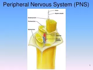
Structure of a Nerve • Nerve – peripheral axons enclosed by connective tissue • Connective tissue coverings include: • Endoneurium – loose connective tissue that surrounds axons • Perineurium – coarse connective tissue that bundles fibers into fascicles • Epineurium – tough fibrous sheath around a nerve
Structure of a Nerve Figure 13.3b
Classification of Nerves • Sensory (afferent) – carry impulse to the CNS • Motor (efferent) – carry impulses from CNS • Mixed nerves – carry somatic and autonomic (visceral) impulses • Most common type
Peripheral Nerves • The four types of mixed nerves are: • Somatic • Sensory • Motor • Visceral • Sensory • Motor • Peripheral nerves can be cranial or spinal
Regeneration of Nerve Fibers • Mature neurons are amitotic • If the soma remains intact, damage can be repaired • Steps • Separated ends seal themselves • Wallerian degeneration of the distal axon by macrophages • Formation of a regeneration tube by the Schwann cell • Guide the axon growth distally
Regeneration of Nerve Fibers Figure 13.4
Regeneration of Nerve Fibers Figure 13.4
Cranial Nerves • Twelve pairs of cranial nerves arise from the brain • They have sensory, motor, or both sensory and motor (mixed nerves) functions • Each nerve is identified by a number (I through XII) and a name
Cranial Nerves Figure 13.5a
Summary of Function of Cranial Nerves Figure 13.5b
Cranial Nerve I: Olfactory • Arises from the olfactory epithelium • Passes through the cribriform plate of the ethmoid bone • Fibers run through the olfactory bulb and terminate in the primary olfactory cortex • Function is the sense of smell
Cranial Nerve I: Olfactory Figure I from Table 13.2
Cranial Nerve II: Optic • Arises from the retina of the eye • Optic nerves pass through the optic canals and converge at the optic chiasm • They continue to the thalamus where they synapse • From there, the optic radiation fibers run to the visual cortex • Functions carry impulses for vision
Cranial Nerve II: Optic Figure II from Table 13.2
Cranial Nerve III: Oculomotor • Motor for movements of the eyes • Parasympathetic fibers innervate the intrinsic muscles of the eye • Constricting the iris, and controlling lens shape
Cranial Nerve III: Oculomotor Figure III from Table 13.2
Cranial Nerve IV: Trochlear Figure IV from Table 13.2
Cranial Nerve V: Trigeminal • Three divisions: ophthalmic (V1), maxillary (V2), and mandibular (V3) • Conveys sensory impulses from various areas of the face (V1) and (V2), and supplies motor fibers (V3) for mastication
Cranial Nerve V: Trigeminal Figure V from Table 13.2
Cranial Nerve VI: Abducens • Primarily a somatic motor nerve Figure VI from Table 13.2
Cranial Nerve VII: Facial • Somatic Motor to the muscles of facial expression, and the transmittal of • Visceral motor to lacrimal and salivary glands • Sensory function is taste from the anterior two-thirds of the tongue
Cranial Nerve VII: Facial Figure VII from Table 13.2