Download
1 / 40
400 likes | 770 Vues
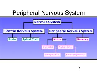
Peripheral Nervous System (PNS). Part 3: Integration & Control. Peripheral Nervous System (PNS). Peripheral Nervous System: All nervous tissue located outside the brain & spinal cord. Components: Cranial Nerves Spinal Nerves Autonomic Nervous System. Subdivisions of the PNS.

E N D
Peripheral Nervous System (PNS) Part 3: Integration & Control
Peripheral Nervous System (PNS) • Peripheral Nervous System: All nervous tissue located outside the brain & spinal cord. • Components: • Cranial Nerves • Spinal Nerves • Autonomic Nervous System
Subdivisions of the PNS • Afferent: The subdivision in charge of sensory information. • Somatic Sensory Division: Carries signals from the skin, muscle, bone, and joint receptors. • Visceral Sensory Division: Carries signals from the organs and tissue in the thoracic & abdominal cavities.
Subdivisions of the PNS • Efferent: The subdivision in charge of motor information. • Somatic Motor Division: Carries signals from the CNS to the skeletal muscles. • Visceral Motor Division aka the Autonomic Nervous System: Controls the part of the body we do not have conscious control over. • Sympathetic Division: “Fight or flight” • Parasympathetic Division: Back to homeostasis!
Types of Nerves in the PNS • Sensory Neurons aka Afferent Neurons: Contain sensory fibers responsible for communicating information about stimuli. • Motor Neurons aka Efferent Neurons: Contain motor fibers responsible for relaying information to the muscular system. • Mixed Neurons: Contain both sensory and motor fibers.
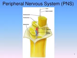
The Nerve Cell • 3 Basic Parts: • Dendrites: Receive information • Soma: Cell body. • Axon: Send information. • May be myelinated or unmyelinated.
Nerve Cells • Ganglion: A cluster of soma’s contained within the same epineurium tissue sheath. • Dorsal Root Ganglia: Contain the somas of the sensory neurons that send impulses to the CNS. • Nerves lying lateral and dorsal to the spinal cord.
Specific Nerve Locations • Dermatomes: Zones in the skin that are served by specific spinal nerves. • In the face & scalp, these are zones served by Cranial Nerve V (Trigeminal). • Muscle Spindles: Contain receptors to allow for stretching of the muscle and knowledge of how the muscle is moving in space. • Intrafusal Muscle Fibers: Nerve endings wrapped around specialized muscle fibers that allows the stretching reflex. • Gamma Motor Neurons: Allow the body to know how far the muscle has stretched. • Proprioceptors: Receptors in muscles and joints that provide knowledge of where the limbs are in space.
Cranial Nerves • Cranial Nerves: Located on the undersurface of the brain. • 24 total held within 12 pairs. • YOU NEED TO KNOW the name, number, type, and function of each nerve!
Cranial Nerves • Cranial Nerve 1: The Olfactory Nerve • Classified as a sensory neuron • Sensory functions include the sense of smell.
Cranial Nerves • Cranial Nerve 2: The Optic Nerve • Classified as a sensory neuron • Sensory functions include the sense of vision.
Cranial Nerves • Cranial Nerve 3: The Oculomotor Nerve • Classified as a motor neuron, but has some sensory functions. • Sensory functions include proprioception (where the body is in space). • Motor functions include moving the eyelids & eye muscles. • Autonomic nervous system functions include the moving the lens and the constriction of the pupil.
Cranial Nerves • Cranial Nerve 4: The Trochlear Nerve • Classified as a motor neuron, but serves some sensory functions. • Sensory functions include propriception (where the body is in space). • Motor functions include moving the eye muscles.
Cranial Nerves • Cranial Nerve 5: The Trigeminal Nerve • Classified as a mixed nerve, serving both autonomic nervous system and sensory functions. • Sensory functions include touch, pain, temperature sensation in face and mouth. • Autonomic nervous system functions include operating the chewing muscles. • 3 Branches of Cranial Nerve 5: • Ophthalmic Nerve: Enters the orbit through the superior orbital fissures; the smallest branch. • Maxillary Nerve: Enters the foramen rotundum; intermediate size. • Mandibular Nerve: Exits through the foramen ovale; the largest branch.
Cranial Nerves • Cranial Nerve 6: The Abducens Nerve • Classified as a motor neuron but serves some sensory functions. • Sensory functions include proprioception (where the body is in space). • Motor functions include moving the eye muscles.
Cranial Nerves • Cranial Nerve 7: The Facial Nerve • Classified as a mixed neuron and serves sensory, motor, and autonomic nervous functions. • Sensory functions include proprioception (where the body is in space) and taste. • Motor functions include the creation of facial expressions. • Autonomic nervous system functions include triggering the secretion of saliva and tears.
Cranial Nerves • Cranial Nerve 8: Vestibulocochlear Nerve aka Auditory Nerve • Classified as a sensory nerve but serves sensory, motor, and autonomic nervous system functions. • Sensory functions include hearing and balance. • Motor functions include modifying inner ear hair cells in response to sound waves. • Autonomic nervous system functions include two branches: • Vestibular Nerve: Carries impulses for equilibrium. • Cochlear Nerve: Carries impulses for hearing.
Cranial Nerves • Cranial Nerve 9: Glossopharyngeal Nerve • Classified as a mixed neuron and serves sensory, motor, and autonomic nervous system functions. • Sensory functions include taste sensations from the tongue and pharynx. • Motor functions include swallowing and speech production. • Autonomic nervous system functions include saliva secretion.
Cranial Nerves • Cranial Nerve 10: The Vagus Nerve • Classified as a mixed neuron, serving sensory, motor, and autonomic nervous system functions. • Sensory functions include taste, blood pressure, breathing & heart rate, and visceral sensations. • Motor functions include swallowing, coughing, and speech production. • Autonomic nervous functions include being the main nerve in control of the autonomic nervous system, particularly the parasympathetic division. • This is the only cranial nerve to leave the head & neck area; innervates the viscera
Cranial Nerves • Cranial Nerve 11: Accessory Nerve • Classified as a motor neuron but also serves sensory and autonomic nervous system functions. • Sensory functions include proprioception (where the body is in space) and voice production. • Motor functions include swallowing and the movement of the head and shoulders. • Autonomic nervous system functions include control of the muscles in the pharynx and larynx.
Cranial Nerves • Cranial Nerve 12: The Hypoglossal Nerve • Classified as a motor neuron but also serves some sensory functions. • Sensory functions include proprioception (where the body is in space). • Motor functions include movement of the tongue.
Autonomic Nervous System (ANS) • Autonomic Nervous System: Controls vital functions that are not under conscious control. • Serves the viscera, including the glands, smooth muscle and cardiac muscle. • Main function is to regulate & maintain homeostasis. • Contracts & relaxes smooth muscle • Increases & decreases rate of contraction in cardiac muscle • Increases & decreases gland secretions.
Types of Neurons in the ANS • ANS Neurons: Instead of a single motor neuron, ANS uses two neurons. • Signals start at a source point in the brain, such as the hypothalamus.
Neurons in the ANS • Autonomic Ganglion: The point at which the neurons diverge. • Preganglionic Neuron: The first neuron in the pair. • Also release acetylcholine (ACh) to stimulate the postganglionic neuron. • Postganglionic Neuron: The second neuron in the pair that leads to the effector neuron. • Secretes noradrenaline (norepinepherine). • Principle of Mass Activation: One preganglionic neuron can excite a large number of postganglionic fibers.
Subdivisions of the ANS • Two Subdivisions of the ANS: • Sympathetic Division aka Sympathetic Nervous System: Initiates the “fight or flight” response mechanism during stress or danger. • Parasympathetic Division aka Parasympathetic Nervous System: Reverses the effects of the sympathetic nervous system to return the body to homeostasis. • Dual Innervation: Concept that most visceral organs are supplied by nerves from both subdivisions. • Allows for cooperative & antagonistic effects.
Sympathetic Division • Sympathetic Division aka Sympathetic Nervous System aka Thoracolumbar Division • Spinal nerves included: Thoracic (T1-12) and Lumbar (L1-2) • Paravertebral Gangli: Sympathetic ganglia located close to the vertebral column. • Preganglionic nerves are much shorter than the postgangionic nerves that must stretch all the way to their target. • Connects to the spinal nerve via both… • White Communicating Rami: Myelinated axons. • Gray Communicating Rami: Unmyelinated axons.
Sympathetic Division • Fight or Flight Mechanism: The body’s method of preparing for physical or emotional stress. • Triggered by noradrenaline release.. Causes: • Simulates rate & force of cardiac muscle contractions • Raises blood pressure • Dilates pupils to allow more light & better vision • Dilates trachea & bronchi to aid breathing • Stimulates liver conversion of glycogen to glucose for energy • Constricts blood vessels in the viscera & skin • Dilates blood vessels in the skeletal muscles, heart, & brain • Slows digestion & urine production
Sympathetic Division • Adrenal Glands: Produce epinephrine (adrenaline) & norepinephrine (noradrenaline) to enhance sympathetic postganglia activity. • Pyramid-shaped glands Located on top of each kidney. • Includes an inner medulla containing modified sympathetic neurons.
Parasympathetic Division • Parasympathetic Division aka Parasympathetic Nervous System aka Craniosacral Division • Contains Cranial Nerves 3, 7, 9, & 10 • Contains Sacral Spinal Nerves (S2-4). • Effector: The target muscle or gland for each postganglionic neuron. • Synapse between preganglionic & postganglionic neurons occur near the effector, leaving postganglionic neurons shorter than the preganglionic • Oppoisite of sympathetic division!
Parasympathetic Division • Parasympathetic stimulation causes: • Slowing of the heart rate to normal rates • Lowers blood pressure to normal rates • Constricts the pupils to normal dilation • Triggers bronchoconstriction (constricting of the airways) to a normal dilation • Increases to normal levels… • Salivation • Lacrimation • Urination • Digestion • Defecation
Good to know about ANS • While ANS is under involuntary control, the parasympathetic division can be manipulated using relaxation techniques. • Examples: Yoga, Buddhist meditation, etc. • Lowers heart rate & oxygen intake far past normal changes of sleep or relaxation!
Cholinergic Receptors • Cholinergic Receptors: Neurons that release acetylcholine (ACh). • Two kinds of receptors on the postsynaptic membrane that bind to ACh: • Nicotinic Receptors: Occur in sympathetic & parasympathetic postganglionic neurons and motor end plates at neuromuscular junctions. • Muscarinic Receptors: Found in all effectors stimulated by the parasympathetic system. • Includes smooth muscle, cardiac muscle, & glands.
Adrenergic Receptors • Adrenergic Receptors: Release norepinephrine (NE). • Make up the majority of sympathetic postganglionic neurons. • Norepinephrine tends to linger longer in the synapses longer than acetylcholine. • Causes effects of adrenergic neurons to last longer.
CNS Control of the ANS • CNS can have a hand in controlling the ANS! • Hypothalamus: Integrates & regulates ANS function. Includes cardiac & pulmonary function, sweating, vasodilation & constriction. • Reticular Formation: Contains centers for cardiac, respiratory, vasomotor, & gastrointestinal functions. • Spinal Cord: Contains centers that regulate the excretory system, without brain involvement.
Side by Side: Sympathetic & Parasympathetic Functions Sympathetic Parasympathetic Inhibits digestion Promotes digestion Increases glycogenolysis Increases glycogen (converts glycogen to synthesis glucose) Increases heart rate Decreases heart rate & force & force Dilates pupils Constricts pupils Dilates airways Constricts airways Stops peristalsis (GI Reengages peristalsis movement) Contracts sphincters Relaxes sphincters
Side by Side: Sympathetic & Parasympathetic Functions Sympathetic Parasympathetic Contracts arrector pili Relaxes arrector pili muscles muscles Triggers vasodilation in… Triggers skeletal muscles vasoconstriction or cardiac muscles vasodilation back to liver normal ranges Triggers vasoconstriction in… kidneys GI tract Increases sweating Causes uterine contraction if pregnant Discharges all stored blood into circulation