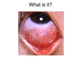
What is it?
What is it?. Conjunctivitis (general). Inflammation of conjunctivae, can be associated with lid inflammation (blepharoconjunctivitis) and corneal inflammation (keratoconjunctivitis).

What is it?
E N D
Presentation Transcript
Conjunctivitis (general) • Inflammation of conjunctivae, can be associated with lid inflammation (blepharoconjunctivitis) and corneal inflammation (keratoconjunctivitis). • Wear gloves. Examine for papillae (bulges, cobblestones) and follicles (grains of rice). Papillae / bacteria. Follicles / viruses. Check for pre-auricular LN. Check acuity. Stain the cornea. • Think of other causes if severe pain or reduced acuity. Photophobia suggests corneal involvement. • Refer if neonatal, follicular, severe purulent discharge, uncertain cause or not responding to treatment.
Viral conjunctivitis • Adenovirus (common). • Highly contagious. Usually bilateral after a few days. Infectious for 2 weeks. Many serotypes but 2 main clinical pictures: • Pharyngoconjunctival fever. Children more likely affected. Associated with viral URTI. Airborne transmission. 30% have mild corneal involvement. • Epidemic keratoconjunctivitis. Contact transmission. 80% have corneal involvement, can be severe. • Management: avoid CL’s, cool compresses, lubricants, hygiene measures. Can take 2-3 wks to resolve, but return if no improvement after 1-2 wks. • No need for exclusion unless there’s an outbreak. BUT in reality most establishments do. • Refer if corneal involvement.
Viral conjunctivitis cont. • HSV: reactivation of HSV-1, in trigeminal ganglia in adults. HSV-2 from vaginal delivery in neonates. • Unilateral, burning pain, FB sensation. Vision blurred if corneal ulceration (typically dendritic). • If in any doubt about corneal involvement, refer. Keratitis can cause scarring and permanently affect vision. • Only manage in GP if no corneal involvement (and no Hx of corneal involvement). Avoid CL’s. Topical aciclovir.
Viral conjunctivitis cont. • Herpes Zoster: reactivation of varicella. Mainly older adults. Hutchinson’s sign indicates high likelihood of eye involvement. Systemic anti-virals ASAP and refer. Lots of complications.
Molluscum contagiosum • Usually in immunocompromised. Look for typical umbilicated nodules. Usually follicular conjunctivitis. Refer for removal.
Bacterial Conjunctivitis • Common, usually benign and self limiting • Children bacterial > viral. Adults viral > bacterial. • Common pathogens: staph, strep. pneumoniae, haemophilus, moraxella. Think about STIs. • Thick mucopurulent discharge, helps to distinguish from viral. Swab if there’s a lot. • Complications rare. Think CL wearers. Otitis media (haemophilus). Chlamydia chronic infection.
Bacterial conjunctivitis cont. • Avoid CL’s. Lid hygiene TDS. Should improve in 10-14 days. • Unclear benefit of Abx. Over prescribed. Don’t alleviate severity of Sx but reduce duration a bit. Ideally wait 7 days, then treat if not better. Continue for 48hrs after resolution. • Chloramphenicol; Fusidic acid. Drops - day. Ointment - night.
Weird and Wonderful conjunctivitis • Contact with pubic lice (mechanical removal plus topical treatment) • Cicatricial diseases • Floppy eyelid syndrome (nocturnal ectropion, associated with obesity and OSA) • Giant papillary conjunctivitis (prolonged contact lens wearing or prostheses, sutures etc). • Superior limbic keratoconjunctivitis. Uncommon. Chronic, intermittent. Middle aged women with thyroid disorders.
Ophthalmia neonatorum • Occurs in first 28 days. • Usually benign. Commonest cause = blocked duct. No longer notifiable. • Pathogens include chlamydia (commonest), gonorrhoea, Staph aureus, strep pneumoniae etc • Less commonly viral or toxic. • Complications can be severe, including death. Refer eye clinic if any doubt.
Blepharitis • Common. Causes sore eyelids. Associated with dry eyes, rosacea, seborrhoeic dermatitis. • Complications rare (include meibomian cysts, styes, entropions, ectropions, corneal scarring, conjunctivitis). • Vicious cycles common (itch, scratch etc) • Three types (often co-exist): staphylococcal, seborroeic, meibomian gland dysfunction.
Seborrhoeic blepharitis • Staphlococcal: ? Why certain people get it. Scaling of lid margins. • Seborrhoeic: Skin is more oily and flakey. ? Reaction to Malassesia Furfur. Associated with rosacea. May benefit from 3 months tetracyclines
Meibomian Gland Dysfunction • Meibomian glands line the lid margins, produce oil that helps tear film to adhere to cornea. • Can become blocked. • Dry eyes, watery eyes. • Lid hygiene - warm, massage, clean.