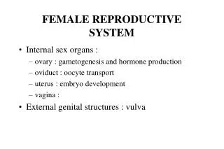
Female reproductive system
330 likes | 610 Vues
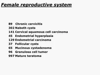
Female reproductive system. 89 Chronic cervicitis 302 Naboth cysts 141 Cervical squamous cell carcinoma 45 Endometrial hyperplasia 129 Endometrial carcinoma 37 Follicular cysts 65 Mucinous cystadenoma 96 Granulosa cell tumor 997 Mature teratoma. Female reproductive system. flashback.

Female reproductive system
E N D
Presentation Transcript
Female reproductive system 89 Chronic cervicitis 302 Naboth cysts 141 Cervical squamous cell carcinoma 45 Endometrial hyperplasia 129 Endometrial carcinoma 37 Follicular cysts 65 Mucinous cystadenoma 96 Granulosa cell tumor 997 Mature teratoma
Female reproductive system flashback 286 CIN (slides with CIN I and CIN II) 90 CIN III 61Leiomyoma 62Leiomyosarcoma
Leiomyoma flashback
Leiomyosarcoma flashback
Chronic cervicitis - this process is invariably associated with an inflammatory infiltrate composed of a mixture of polymorphonuclear leukocytes and mononuclear cells, and if the inflammation is severe, it may be associated with loss of the epithelial lining (erosion or ulceration) and epithelial repair - reserve cells in the transformation zone may undergo columnar or squamous differentiation (metaplasia)
Naboth cysts during the squamous metaplasia (the process of transformation from a columnar to a squamous lining),the squamous epithelium can overgrow and obliterate the surface columnar papillae, so it covers and obstructs crypt openings, with the accumulation of mucus in deeper crypts (glands) to form mucous (nabothian) cysts
Cervical epithelium without CIN flashback
CIN flashback
CIN III flashback
Cervical squamous cell carcinoma - irregularly shaped nests and clusters of squamous cells invading stroma, often with eosinophilic cytoplasm, atypical nuclei with coarse chromatin, prominent nucleoli, and abnormal mitotic figures - keratinizing or non-keratinizing
Endometrial hyperplasia Simple hyperplasia -glands cystically dilated, with occasional outpouchings surrounded by abundant cellular stroma - sometimes glands only minimally dilated but focally crowded - cells lining glands are pseudostratified and columnar and cytologic atypia is absent. Complex hyperplasia - crowded glands with little intervening stroma - back-to-back glands with papillary intraluminal infoldings - usually gland outlines highly complex but can be tubular - epithelial stratification usually prominent
Endometrial carcinoma - neoplastic glands resembling proliferative-type endometrial glands, with nuclear stratification, cribriform glandular patterns, papillary growth patterns, and evidence of stromal invasion - grading is based on architectural pattern and nuclear features. Architectural grade is determined by the extent to which the tumor is composed of solid masses of cells as compared with well-defined glands
Mucinous cystadenoma - cysts lined by a single layer of non-stratified columnar mucinous epithelium that is either of intestinal type or endocervical type
Follicular cysts - usually multiple - range in size up to 2 cm in diameter - filled with a clear serous fluid - granulosa lining cells can be identified histologically if the intraluminal pressure has not been too great - the outer theca cells may be conspicuous with increased cytoplasm and a pale appearance (luteinized)
Granulosa cell tumor - variety of patterns, including microfollicular, macrofollicular, trabecular, solid-tubular, gyriform, and diffuse (sarcomatoid) - better differentiated forms show follicular structures composed of granulosa cells arranged around a central eosinophilic hyaline or fibrillar material (Call-Exner bodies) - the typical cytologic feature is a longitudinal nuclear groove giving the nucleus the appearance of a coffee bean - mitotic activity can be rare to abundant
Mature teratoma - cystic structure lined predominantly by skin and cutaneous adnexal structures, usually with abundant sebaceous and sweat glands - hair almost always present - other components include cartilage, bone, bronchial or gastrointestinal epithelium, and mature glial tissue - if only skin and adnexal structures present can be termed dermoid cyst - thyroid tissue can be found in up to 20% of cases (when thyroid tissue is the predominant tissue the lesion is called struma ovarii)