Download
1 / 21
260 likes | 676 Vues
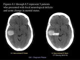
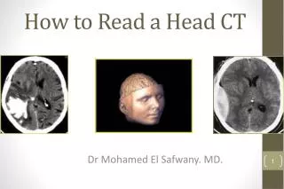
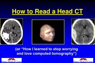
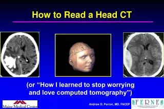
Systematic Approach to Reading a Non-Contrast Head CT Scan. Systematic Approach. Assess each component of the intracranial cavity Blood Brain Bone CSF. Blood. Epidural Hematoma (EDH) Arterial Bleed: High-Pressure “Lens” Shape Middle Meningeal Laceration (Skull Fracture)

E N D
Systematic Approach Assess each component of the intracranial cavity • Blood • Brain • Bone • CSF
Blood • Epidural Hematoma (EDH) Arterial Bleed: High-Pressure “Lens” Shape Middle Meningeal Laceration (Skull Fracture) • Subdural Hematoma (SDH) Venous Bleed: Low-Pressure “Crescent” Shape Acute / Chronic (Is Patient Anticoagulated? Alcoholic?) • Subarachnoid Hemorrhage (SAH) Traumatic / Aneurysmal • Intraventricular Hemorrhage (IVH) • Intraparenchymal Hemorrhage (IPH) Hypertensive Basal Ganglia or Lobar Cerebral Contusion “Coup-Contrecoup”
Contusion Coup (Red) Contrecoup Contusion (Orange) (at 180 degrees)
Brain • Symmetry (of Hemispheres) Hyper-/Hypodensities (Masses, Edema, Stroke) • Grey/White Matter Border Lost Grey-White Differentiation in Anoxic Injury • Midline Shift • Gyri/Sulci Wide Sulci: Atrophy Effaced Sulci: Edematous “Tight/Swollen” Brain • Pneumocephalus (Air in Brain) Open Fracture (or Craniotomy) Fracture through Sinus
Bone • Fractures Especially Temporal Bones • Sinuses & Air Cells Look for Air-Fluid Levels
CSF: Ventricles & Cisterns • Ventricles Blood in Ventricles? Effacement/Asymmetry: Compression from Mass/Hematoma Hydrocephalus Atrophy (“Ex Vacuo”) Communicating / Obstructive Hydrocephalus If Obstructive, Where? Look at Lateral, 3rd, and 4th Ventricles • Cisterns Look for Effacement (Edema/Early Herniation) Look for Blood