Download
1 / 19
190 likes | 420 Vues
Research course on functional magnetic resonance imaging (non-invasive brain imaging) . Juha Salmitaival. Outline. MRI safety course Introductory lectures ( next week F227!) Scanning (2 sessions for each participant ) Preprocessing Data-analysis Writing a research report

E N D
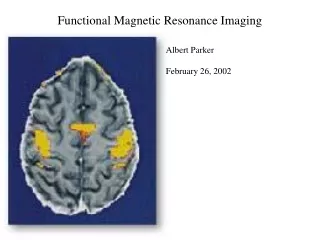
Research course on functional magnetic resonance imaging (non-invasive brain imaging) Juha Salmitaival
Outline • MRI safetycourse • Introductorylectures (nextweek F227!) • Scanning (2 sessions for eachparticipant) • Preprocessing • Data-analysis • Writing a researchreport • Note! Youwillnotbeable to plan and prepare the studiesyourselves -> 5 cr
Today’slecture • Overview of the stages in an fMRIstudy • MRI signal • BOLD hemodynamics & physiology • MRI protocol • Scanningsettings • MRI images • Someartefacts • FSL introduction • Brainextraction
Stages of an fMRIstudy • Researchplan, funding • Ethicalpermission (HUCH) and researchpermission (AMI centre) • Settingup the experiment (stimulation, MRI protocol) and piloting • Collecting the data • Data-analysis • Preprocessing (motioncorrection, spatial/temporalfiltering, brainextraction) • Data-analysis (model-based, e.g., GLM, data-driven, e.g., ICA, ISC) • Writing a manuscript
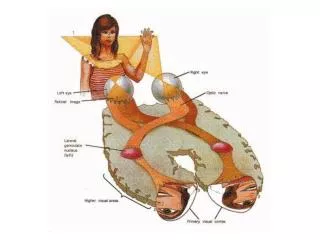
MRI signal • B0 field (e.g., 3T) • Larmorfrequency • RF excitation / relaxation • T1 = realignmentwith the magneticfield • T2 = emission of energy • T2* = sensitive to inhomogeneties in the magneticfield
MRI signal • Summary of MRI • Gradientfield • MORE INFORMATION: • http://www.cis.rit.edu/htbooks/mri/
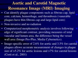
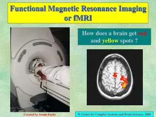
BOLD hemodynamics • BOLD (bloodoxygenationdependent)signal • Ittakesabout 4-6 seconds to reachitspeak • HRF variesbetweensubjects and brainregion
BOLD physiology • Neuronalactivity • Energy consumption LFP and BOLD • Metabolicpathway (local)
MRI protocol • MRI sequence (RF excitation, gradientpulses) • Localizer, epi-sequence, anatomicalsequence • TR (1.5 – 4 sec.), slicethickness (2-5 mm), number of slices (1-50), aquisitionmatrix (64 x 64 – 192 x 192), FOV, number of samples • Continuousimaging (jitter?) orsparsetemporalsampling
Scanning ”settings” • Fatsuppression • Spectralspatial RF pulseminimumslicethickeness 3mm • Spectral RF (slicethickness < 3 mm) • Shimming (fMRIautoshim, DTI HOS - manual) • Optimizing the homogeneity of the B0 field • Correction of the inhomogeneitycanalsobedone • Prescan (use auto prescan) • Optimalresonancefrequency, adjustingtransmit and receivergain
MRI image • Voxel (pixel in 3d) • Slicethickness x FOV/matrix x FOV/matrix (in-planeresolution) • Volume (sample) • E.g., 30 x 64 x 64 • 4d image (typically > 100 MB, < 2 GB) • Formats: dicom, analyze, nifti, niftigz
Anatomical and slicedirections • Anatomicaldirections • Superior-inferior (head-foot) • Anterior-posterior (front-back) • Dorsal-ventral (back-front) • Right-left • Slicedirections • Axial • Coronal • Sagittal
Artefacts (some of those) • Movement • Cross-talk • Aliasing • Chemicalshift • Susceptibilityartefact • Nyquist ghosting • Geometricdistortion
Imagepreparation • Dicom2nifti conversion (dcm2niigui) • http://www.cabiatl.com/mricro/mricron/install.html • Output: FSL (4D NifTI) orCompressed FSL • Imageviewing • Fslview (http://www.fmrib.ox.ac.uk/fsl/fslview/index.html) • MRIcron (http://www.cabiatl.com/mricro/mricron/index.html) • Data check • Orientation, artefacts
Toolboxselection • Stimulus presentation • Presentation (nbs) • E-prime • Matlab • Data-analysis • FSL • SPM • Brainvoyager • Freesurfer • AFNI • GIFT • Remember to add FSL to yourbash
Homework - FSL Introduction • Website (www.fmrib.ox.ac.uk/fsl/fsl/list.html) • FSLUTILS • fslinfo • fslmaths • BET • FLIRT/FNIRT • FEAT • MELODIC
Brainextraction • Needed for imageregistration and artifactrejection
References & Images • FSL-course • http://www.fmrib.ox.ac.uk/fslcourse/ • SPM-course • http://www.fil.ion.ucl.ac.uk/spm/course/