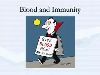
Understanding Immunity: Cells and Defenses
410 likes | 510 Vues
Learn about the key components of immunity, including lymphocytes, innate immunity, and natural killer cells. Explore how the immune system adapts to fight infections and provides defense against pathogens. Discover the roles of sentinel cells in triggering immune responses.

Understanding Immunity: Cells and Defenses
E N D
Presentation Transcript
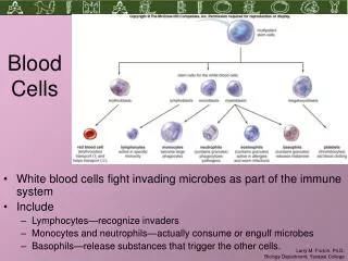
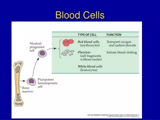
Lymphocytes http://www.vet.uga.edu/IVCVM/1998/latimer1/latimer1.htm
In biology, immunity is the state of having sufficient biological defences to avoid viral, bacterial, fungal infection, or other unwanted biological invasion. • Immunity involves both specific and non-specific components. • The non-specific components act either as barriers or as eliminators of wide range of pathogens irrespective of antigenic specificity. • Other components of the immune system adapt themselves to each new disease encountered and are able to generate pathogen-specific immunity.
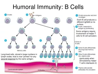
A further subdivision of adaptive immunity is characterized by the cells involved; humoral immunity is the aspect of immunity that is mediated by secreted antibodies. • The protection provided by cell mediated immunity involves T-lymphocytes alone. • Humoral immunity is active when the organism generates its own antibodies, and passive when antibodies are transferred between individuals. • Similarly, cell mediated immunity is active when the organisms’ own T-cells are stimulated and passive when T cells come from another organism.
Innate immune system • Innate immunity, or nonspecific immunity is the natural resistances and provides resistances through several physical, chemical and cellular approaches. • Microbes first encounter the epithelial layers, physical barriers that line skin and mucous membranes. • Subsequent general defences include secreted chemical signals (cytokines), antimicrobial substances, fever, and phagocytic activity associated with the inflammatory responses. • The phagocytes express cell surface receptors that can bind and respond to common molecular patterns expressed on the surface of invading microbes.
Innate immune system • The innate immune system, comprises the cells and mechanisms that defend the host from infection by other organisms in a non-specific manner. • Cells of the innate system recognize and respond to pathogens in a generic way. • Unlike the adaptive immune system (which is only found in vertebrates), it does not confer long-lasting or protective immunity to the host. • Innate immune systems provide immediate defense against infection, and are found in all classes of plant and animal life. • The innate immune system is an evolutionarily older defense strategy, and is the dominant immune system found in plants, fungi, insects, and in primitive multicellular organisms.[ http://en.wikipedia.org/wiki/Innate_immunity
The major functions of the vertebrate innate immune system include: • Recruiting immune cells to sites of infection, through the production of chemical factors, including specialized chemical mediators, called cytokines. • Activation of the complement cascade to identify bacteria, activate cells and to promote clearance of dead cells or antibody complexes. • The identification and removal of foreign substances present in organs, tissues, the blood and lymph, by specialised white blood cells. • Activation of the adaptive immune system through a process known as antigen presentation. • Acting as a physical and chemical barrier to infectious agents.
Natural killer cells • Natural killer cells (or NK cells) are a type of cytotoxiclymphocyte critical to the innate immune system. • NK cells provide rapid responses to virally infected cells and respond to tumor formation, acting at around 3 days after infection. • Typically immune cells detect MHC presented on infected cell surfaces, triggering cytokine release, causing lysis or apoptosis. • NK cells are unique, however, as they have the ability to recognize stressed cells in the absence of antibodies and MHC, allowing for a much faster immune reaction. • They were named “natural killers” because of the initial notion that they do not require activation in order to kill cells that are missing “self” markers of major histocompatibility complex (MHC) class 1. http://www.youtube.com/watch?v=HNP1EAYLhOs http://en.wikipedia.org/wiki/Natural_killer_cell
http://upload.wikimedia.org/wikipedia/commons/a/a9/Innate_immune_system.pnghttp://upload.wikimedia.org/wikipedia/commons/a/a9/Innate_immune_system.png
Sentinel cells - a very general term for any cell type that plays a prominent role in host defense by sensing and monitoring the presence of foreign antigens. Processing of these antigens and recognition by appropriate immune effector cells such as B-cells and T-cells eventually initiates humoral and cell-mediated immune responses. • The term comprises specialized cell types such as antigen-presenting cells, macrophages, mast cells, dendritic cells, Langerhans cells and also includes specialized T-cell populations. • The term may include also fibroblasts, epithelial cells (for example, mesothelial cells) and other cell types that may not primarily process and present antigens to other immune cells. Such cells are important for innate immunity because they can release different patterns of chemokines and cytokines that attract inflammatory cells and immune cells able to trigger immune responses. • The term sentinel cells has been used also for a variety of neurocrine and endocrine cell types that maintain homeostasis in different biological systems (e. g., glucose homeostasis). http://www.copewithcytokines.de/cope.cgi?key=sentinel%20cells
Adaptive imunity • The adaptive immune system, which has been best studied in mammals, originated in a jawed fish approximately 500 million years ago. Most of the molecules, cells, tissues, and associated mechanisms of this system of defense are found cartilaginous fishes. • Lymphocyte receptors, Ig and TCR, are found in all jawed vertebrates. • The most ancient Ig class, IgM, is membrane-bound and then secreted upon stimulation of cartilaginous fish B cells. • Another isotype, shark IgW, is related to mammalian IgD. • TCRs, both α/β and γ/δ, are found in all animals from gnathostomes to mammals. • Like TCR and Ig, the MHC is found only in jawed vertebrates. • Genes involved in antigen processing and presentation, as well as the class I and class II genes, are closely linked within the MHC of almost all studied species.
Adaptive imunity • Adaptive immunity is often sub-divided into two major types depending on how the immunity was introduced. • Naturally acquired immunity occurs through contact with a disease causing agent, when the contact was not deliberate. • Artificially acquired immunity develops only through deliberate actions such as vaccination. • Both naturally and artificially acquired immunity can be further subdivided depending on whether immunity is induced in the host or passively transferred from an immune host. • Passive immunity is acquired through transfer of antibodies or activated T-cells from an immune host, and is short lived—usually lasting only a few months. • Active immunity is induced in the host itself by antigen and lasts much longer, sometimes lifelong.
Antigene presenting cell antigene MHC peptide T lymphocyte B lymphocyte lymphokines Activated T lymphocyte Activated B lymphocyte antibodies
B and T cell proliferation Memory B cells Activated B cell Peripheral B cell Lymph follicle Clone Clone CFU B cell CFU Red bone marrow Antigen APC T helper (Th) CFU T cell Plasmocyte Peripheral T cell Activated T cell T suppresor (Ts) Thymus Lymph follicle T cytotoxic (Tc) T killer (Tk) Memory T cells
Antigene presentation • Antigen processing is an immunological process that prepares antigens for presentation to special cells of the immune system called T lymphocytes. • It is considered to be a stage of antigen presentation pathways. This process involves two distinct pathways for processing of antigens from an organism's own (self) proteins or intracellularpathogens (e.g. viruses), or from phagocytosed pathogens (e.g. bacteria); subsequent presentation of these antigens on class I or class II MHC molecules is dependent on which pathway is used. • Both MHC class I and II are required to bind antigen before they are stably expressed on a cell surface. • MHC I antigen presentation typically (considering cross-presentation) involves the endogenous pathway of antigen processing. • MHC II antigen presentation involves the exogenous pathway of antigen processing. • Cross-presentation involves parts of the exogenous and the endogenous pathways but ultimately involves the latter portion of the endogenous pathway (e.g. proteolysis of antigens for binding to MHC I molecules). • While the conventional distinction between the two pathways is useful, there are instances where extracellular-derived peptides are presented in the context of MHC class I and cytosolic peptides are presented in the context of MHC class II (this often happens in dendritic cells). http://www.youtube.com/watch?v=LwLYGTS_3EI http://en.wikipedia.org/wiki/Antigen_processing
The exogenous pathway • The exogenous pathway is utilized by specialized antigen presenting cells to present peptides derived from proteins that the cell has endocytosed. • The peptides are presented on MHC class II molecules. • Proteins are endocytosed and degraded by acid-dependent proteases in endosomes; this process takes about an hour. • The nascent MHC class II protein in the rough ER has its peptide-binding cleft blocked by Ii (the invariant chain; a trimer) to prevent it from binding cellular peptides or peptides from the endogenous pathway. • The invariant chain also facilitates MHC class II's export from the ER in a vesicle. • This fuses with a late endosome containing the endocytosed, degraded proteins. • The invariant chain is then broken down in stages, leaving only a small fragment called CLIP which still blocks the peptide binding cleft. • An MHC class II-like structure, HLA-DM, removes CLIP and replaces it with a peptide from the endosome. • The stable MHC class-II is then presented on the cell surface.
MHC class II heterodimers assemble in the endoplasmic reticulum (ER) with the assistance of invariant chain (Ii). • The cytoplasmic tail of Ii contains a motif that targets the Ii–MHC class II complex to the endosomal pathway, either directly from the trans-Golgi network (TGN) to early endosomes or via the plasma membrane. • Ii–MHC class II complexes at the cell surface are rapidly internalized into recycling endosomes and then traffic to the early endosomes. • Maturation of the early endosome leads to activation of lysosomal enzymes, including cysteine proteases, which degrade endogenous endosomal proteins, internalized proteins and Ii. • In addition to these proteases, lysosomal reductases that cleave disulphide bonds, such as -interferon-inducible lysosomal thiol reductase (GILT), are required for the processing of protein antigens that contain disulphide bonds. • Following Ii cleavage, the MHC class II peptide-binding groove remains occupied by the class-II-associated invariant chain peptide (CLIP), which prevents premature peptide loading. • Removal of CLIP and loading of peptides is mediated by the MHC-like molecule H–2M. • These peptide–MHC class II complexes then traffic to the plasma membrane. TCR, T-cell receptor. Nature Reviews Immunology 3, 472-482 (June 2003)
Invariant chain (Ii) is degraded in a stepwise manner in the endosomes. • The initial cleavage is thought to be mediated by a leupeptin-insensitive cysteine protease or an aspartic protease, whereas subsequent steps are a result of the activity of leupeptin-sensitive cysteine proteases and leave the MHC class II peptide-binding groove occupied by the class-II-associated invariant chain peptide (CLIP). • Aspartic and cysteine proteases also degrade internalized and endogenous proteins that are present in the endosomal compartment, and the MHC-like molecule H–2M then exchanges CLIP for the peptides that are generated by these enzymes. • The size and relative abundance of the Ii fragments that accumulate in the presence of leupeptin vary depending on the species from which the antigen-presenting cell was derived and the MHC haplotype.
The endogenous pathway • The endogenous pathway is used to present cellular peptide fragments on the cell surface on MHC class I molecules. • If a virus had infected the cell, viral peptides would also be presented, allowing the immune system to recognize and kill the infected cell. • Worn out proteins within the cell become ubiquitinated, marking them for proteasome degradation. • Proteasomes break the protein up into peptides that include some around nine amino acids long (suitable for fitting within the peptide binding cleft of MHC class I molecules). • Transporter associated with antigen presenting (TAP), a protein that spans the membrane of the rough endoplasmic reticulum, transports the peptides into the lumen of the rough endoplasmic reticulum (ER). • Also within the rough ER, a series of chaperone proteins, including calnexin, calreticulin, ERp57, and Binding immunoglobulin protein (BiP) facilitates the proper folding of class I MHC and its association with β2 microglobulin. • The partially folded MHC class I molecule then interacts with TAP via tapasin (the complete complex also contains calreticulin and Erp57 and, in mice, calnexin). • Once the peptide is transported into the ER lumen it binds to the cleft of the awaiting MHC class I molecule, stabilizing the MHC and allowing it to be transported to the cell surface by the Golgi apparatus.
Cross-presentation • In Cross-presentation, peptides derived from extracellular proteins are presented in the context of MHC class I. • The cell starts off with the exogenous pathways but diverts the antigens (cytosolic diversion) to the endogenous pathway. • This can allow the cell to skip the parts of the endogenous pathway that involve synthesis of antigens from the antigenic genes with cellular machinery upon infection. • The endogenous pathway can involve infection before being able to present antigens with MHC I, and cross-presentation saves them the effort needed for that and allows the professional antigen-presenting cells (dendritic cells) to process and present antigens without getting infected. • This does not tend to happen to dendritic cells and is quite common scenario of antigen-processing using the endogenous pathway. • Not all cells utilize cross-presentation. If they did, all of them would be involved.MHC I
Pathways to antigen presentation Nature Volume:471,Pages:581–582 Date published: (31 March 2011)
a, Direct presentation occurs when an antigen-presenting cell such as a dendritic cell is infected, and displays processed antigenic peptides in complex with MHC class I molecules on its surface, thereby activating T cells. b, In cross-presentation, dendritic cells acquire antigens obtained by infected cells through endocytosis and phagocytosis, and — with or without some processing — load them onto class I molecules for presentation to T cells. c, In a third pathway, called cross-dressing, dendritic cells acquire preformed MHC class I molecules in complex with antigens from other cells by the process of trogocytosis or through gap junctions. Wakim and Bevan4 show that cross-dressing is used to activate memory T cells, but not naive T cells, in response to viral infection.
All dendritic cells (DCs) have functional MHC class I and MHC class II presentation pathways. • MHC class I molecules present peptides that are derived from proteins degraded mainly in the cytosol, which in most DC types comprise almost exclusively endogenous proteins (synthesized by the cell itself). • MHC class II molecules acquire peptide cargo that is generated by proteolytic degradation in endosomal compartments. • The precursor proteins of these peptides include exogenous material that is endocytosed from the extracellular environment, and also endogenous components, such as plasma membrane proteins, components of the endocytic pathway and cytosolic proteins that access the endosomes by autophagy. • CD8+ DCs have a unique ability to deliver exogenous antigens to the MHC class I (cross-presentation) pathway, although the mechanisms involved in this pathway are still poorly understood. • The bifurcated arrow indicates that the MHC class II and the MHC class I cross-presentation pathways may 'compete' for exogenous antigens in CD8+ DCs, or that the endocytic mechanism involved in internalization of a given antigen may determine whether it is preferentially delivered to the MHC class II pathway or the MHC class I cross-presentation pathway. • TAP, transporter associated with antigen processing.
Role of Cytokines Cytokines are essential in any type of immune response. These are proteins secreted by cells of the immune system to signal other cells of the immune system. Typically they are produced locally and affect cells in the near vicinity. The following table lists some of the cytokines involved in generating an immune response.
Table 1. From Basic Science of Tumor Antigen and Immune activation, Handbook of Cancer Vaccines and http://web.indstate.edu/thcme/mwking/growth-factors.html II. Immune properties of Tumors
Role of CytokinesCytokines are essential in any type of immune response. These are proteins secreted by cells of the immune system to signal other cells of the immune system. Typically they are produced locally and affect cells in the near vicinity. The following table lists some of the cytokines involved in generating an immune response. Table 1. From Basic Science of Tumor Antigen and Immune activation, Handbook of Cancer Vaccines and http://web.indstate.edu/thcme/mwking/growth-factors.html II. Immune properties of Tumors