Blood cells
Blood cells. Types of blood cells. Red blood cells Macrophage system Lymphatic system. Red blood cells. Functions Transport of hemoglobin Oxygen Free hemoglobin can be filtered into the urine by kidney in higher animals Must be in the cell Formation of carbonic acid

Blood cells
E N D
Presentation Transcript
Types of blood cells • Red blood cells • Macrophage system • Lymphatic system
Red blood cells • Functions • Transport of hemoglobin • Oxygen • Free hemoglobin can be filtered into the urine by kidney in higher animals • Must be in the cell • Formation of carbonic acid • Carbonic anhydrase (water plus CO2) • Faster clearance of CO2 from the body • Biological buffer
Shape and size of RBC • Flexible bag • Passing through the capillary • No membrane stretching • Greater membrane to volume ratio • Concentrations • 5,200,000/ml in men and 4,700,000/ml in women (300,000 give or take)
Hemoglobin concentration • 34g/100ml cell (no plasma) • Upper metabolic limit • Almost always around the maximum • Hematocrit (% cell in blood) • 40-45% • 15g/100 ml blood in male and 14g/100 ml blood in female • Each g hemoglobin can carry 1.34 ml oxygen • 20ml O2/100 ml blood in men and 19ml O2/100 ml blood in women
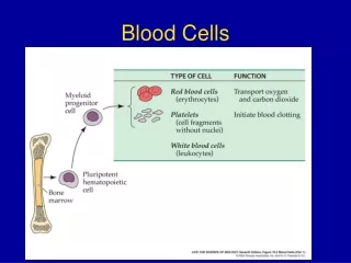
RBC production • Areas of body • Fetal stage • Yolk sac during embryonic development • Liver during middle trimester • Spleen and lymph nodes • Postnatal stage • Bone marrow • Switch during the last month of gestation
RBC production • Areas of body • Adult • Membranous bones • Ability decreases as one ages
Bone marrow Yolk sac Vertebra Liver Sternum Rib Spleen Femur Tibia 20 3 1 ADULT FETAL MONTHS
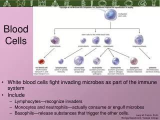
Generation of blood cells • Pluripotent hematopoetic stem cells • Reservoir • Committed hematopoetic stem cells • Committed stem cells • Erythrocyte • Derived from colony forming unit-erythrocytes (CFU-E) • Granulecytes and monocytes • Derived from CFU-GM
Growth and differentiation of stem cells • Growth inducers • Differentiation inducers • Commitment of stem cells to differentiate • Production controlled by external factor • Low blood O2 • Infection (WBC)
Stages of differentiation • Proerythroblast • Basophil erythroblast • Stain with basic dye • Increased % hemoglobin as the stage progresses • Condensation and loss of nucleus and other organelles
Regulation of RBC production • Total mass of RBC in circulation • Narrow range • Adequate # of RBC for O2 transport • No impact on blood flow • Oxygenation of tissue • Most essential regulator • Loss of RBC/loss of O2 carrying capacity
Regulation of RBC production • Erythropoetin • Stimulates RBC production when low O2 states • Kidney • Main source (90%) • Stimulated by low oxygen availability to tubular cells • Production signaled by other parts of body
Regulation of RBC production • Erythropoetin • Rapid production • Maximum within 24 hours after hypoxia • Stimulates proerythroblast production from stem cells • Increased rate of differentiation
RBC maturation • RBC • Most rapidly growing and reproducing cells • Vitamins • Vitamin B12 and folic acid • Synthesis of TTP • Essential for nuclear maturation and cell division • Formation of macrocytes (low O2 carrying capacity) when low
Pernicious anemia • Poor vitamin B12 absorption • Atrophy of GI nucosa that causes loss of intrinsic factor for vitamin B12 absorption • Susceptible to digestion • No interaction with blush border in ileum • Reduced B12 being carried in blood • Needs 3-4 years before the symptom appears • Stored in liver
Anemia caused by folic acid deficiency • Spruce • Small intestine disease that reduce folic acid and vitamin absorption
Hemoglobin formation • Stages • Formation of succinyl-CoA • Krebs cycle • Combination of succinyl-CoA with glycine • Pyrrole • Formation of protoporophyrin • Four pryrroles • Formation of heme • protoporophyrin plus iron • Combination of heme with globulin protein
Types of hemoglobin chains • Four types • Alpha, beta, gamma, and delta • Hemoglobin A = two alpha plus two beta chains • Determines oxygen binding affinity • Sickle cell anemia • Amino acid substitution in beta chains • Combination of O2 with hemoglobin • Loose interaction with coordination bonds of iron atom • Reversible • Carried as O2 rather than oxygen ion
Iron metabolism • Total iron quantity • 4-5 g • 65 % in hemoglobin • Transport and storage • Bound to plasma proteins after absorption • Bound to ferritin in the cell • Storage • Released when plasma concentrations are low
Daily iron loss • 0.6 mg per day • 1.3 mg/day during menstruation • Absorption of iron • Small intestine • Bound to apotransferrin (bile product) to form transferrin • Regulation of total body iron
Life span of RBC • Average life span • 120 days • Metabolically active • Enzymes • Pliability • Iron transport • Iron maintenance • Oxidation prevention • Become fragile • Loss of metabolism
Destruction of RBC • Spleen • Self-destruction through narrower passageway • Structural trabecule of red pulp • Hemoglobin • Phagocytosis (macrophage) • Kupffer cells in liver and spleen • Iron • Recycled • Porphyrin • Converted to bilirubin
Anemia • Hemoglobin deficiency • Blood loss • Very small RBC (microcytic, hypochromic) • Bone marrow aplasia (loss of function) • Vitamin deficiency • Abnormally large RBC (megaloblastic) • Abnormality of RBC (hereditary) • Sickle cell anemia • Erythroblastosis fatalis
Polycythemia • Excess RBC • Hypoxia • Physiologic polycythemia • Low O2 content due to high altitude • Polycythemia Vera • genetic aberration • Increase in blood viscosity • Increased arterial pressure
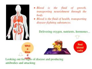
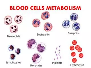
Defense against infection • Leukocytes • White blood cells • Tissue cells • Methods • Phagocytosis • Physical destruction • Antibody production and lymphocyte sensitization
Leukocytes • Bone marrow • Granulocytes • Monocytes • Lymphocytes • Lymph tissue • Lymphocytes • Plasma cells • Mobile unit of defense system
Types • Granular appearance (granulocytes, 65% of total WBC) • Multiple nucleus • Polymorphonuclear neutrophils • Polymorphonuclear eosinophils • Polymorphonuclear basophils
Types • Monocytes (5 %) • Lymphocytes (30 %) • Plasma cells • Platelets • Fragments of megakaryocytes
Granulocytes and monocytes • Phagocytosis • Lymphocytes and plasma cells • Connection with immune system • Platelets • Blood clotting
Genesis of WBC • Pluripotent hematopoietic stem cell • Two lineage for WBC • Myelocytic (myeloblast) • Lymphocytic (lymphoblast) • Site of generation • Bone marrow • Granulocytes and monocytes • Lymph system
Life span • Granulocytes • 4-8 hours after being released in circulation • 4-5 days in tissue • Monocytes • 10-20 hours in circulation • Up to months in tissue • Transformed into macrophage
Neutrophils and macrophages • Initial defense against infection • neutrophils • Active in blood • Macrophage • Exist as monocytes in circulation
Movement of WBC between circulation and tissue • Initiated by chemotaxis • Toxins • Chemicals released from damaged/infected tissue • Complement complex • Diapedesis • Sliding through the pore • Ameboid motion
Phagocytosis • Neutrophils • Mature cells • Phagocytize 3-20 bacteria per cell • No regeneration • Macrophage • Mature monocyte • Must enter the tissue • Phagocytize 100 bacteria/cell
Production of bactericidal agents • Oxidizing agents • Superoxide • Hydrogen peroxide • Hydroxyl ion • Hypochlorite (chloride plus hydrogen peroxide)
Monocyte-macrophage cell system • Present in all tissues • Skin • Lymph nodes • Lung aleveoli (giant cells) • Liver (Kupffer cells) • Spleen • Composition • Monocytes, mobile macrophage, and fixed macrophage
Inflammation • Change of tissues due to injury • Surrounding area by chemicals • Vasodilation (excess local blood flow) • Increased capillary permeability • Clot formation • Granulocyte and monocyte migration • Cell swelling
Removal of damaged tissue by macrophage • Activated by chemical signals • Injuring living tissue by macrophage • Walling off the injured area • Fibrinigen clot to separate injured area from healthy tissue • Intensity of inflammation • Degree of tissue damage
Neutrophil and macrophage response • Tissue macrophage • First line of defense • Enlargement • Mobilization • Migration of neutrophils • Initiated by chemotaxis • Margination (increased stickiness of endotherial surface) • Diapedesis
Increased production of neutrophils • Neutrophilia • Chemical signals • Migration of macrophage • Migration of monocytes • Increased production of granulocytes and monocytes • Formation of pus • Necrotic tissue • Dead neutrophhils and macrophages • Tissue fluid
Eosinophils • Weak phagocytes • Small portion of total leukocytes (2 %) • High in people with parasite infection • Attach themselves onto the parasite and produce chemicals to eliminate paracites • Collect in tissues with allergic reaction • Chemicals from other cells • Prevent spread of allergic inflammation
Basophils • Similar to tissue mast cells • Liberate heparin (anticoagulant) • Release histamine • Small amount of serotonin and bradykinin • Allergic reaction • IgE attach to mast cell/basophils
Abnormalities • Leukopenia • Production of low leukocytes by bone marrow • Very acute • Radiation and drugs • Leukemia • Uncontrolled leukocyte production • Lymphotic or myelogenous leukemia • Release of undifferentiated cells