White blood cells
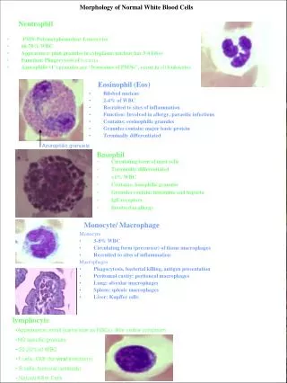
White blood cells. Definition: White blood cells or leukocytes are cells of the immune system which defend the body against both infectous disease and foreign materials. Characters of WBCs :


White blood cells
E N D
Presentation Transcript
White blood cells • Definition: • White blood cells or leukocytesare cells of the immune system which defend the body against both infectous disease and foreign materials. • Characters of WBCs: • Whenever a germ or infection enters the body the white blood cells have a variety of ways by which they can attack. Some will produce protective antibodies that will overpower the germ. Others will surround and devour the bacteria. • The white blood cells have a rather short life cycle, living from a few days to a few weeks. • Several different and diverse types of leukocytes exist, but they are all produced and derived from a multipotent cell in the bone marrow known as a hematopoietic stem cell.
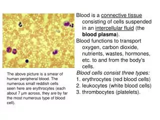
Characters of WBCs • Leukocytes are found throughout the body, including the blood and lymphatic system. • The name "White Blood Cell" derives from the fact that after cenrifugation of a blood sample, the white cells are found in the Buffy coat, a thin layer of nucleated cells between the sedimented red blood cells and the blood plasma, which is typically white in color. The scientific term leukocyte directly reflects this description, derived from Greekleuko - white, and cyte - cell.
White cell count (WBC) • White cell count (WBC) is the total number of leukocytes in a volume of blood, expressed as thousands/µl. • As with the RBC, the WBC can be done by manual methods or by automated cell counters. • Normal Values: • Newborn 9.0-30.0 x 10³/μl • 1 week 5.0-21.0 x 10³/μl • 1 month 5.0-19.5 x 10³/μl • 6-12 months 6.0-17.5 x 10³/μl • 2 years 6.2-17.0 x 10³/μl • Child/adult 4.8-10.8 x 10³/μl
Leukocytosis • Leukocytosis is a condition characterized by an elevated number of white cells in the blood, which is usually due to: • Bacterial infection such as appendicitis, tonsillitis, ulcers and urinary tract infection • Leukemia. • Pregnancy. • Hemolytic disease of new born. • Following exercise. • Emotional stress. • Food intake.
Leukopenia • Leukopenia is a condition characterized by a decreased number of white cells in the blood, which is usually due to: • Viral disease such as measles and infectious hepatitis. • Some bacterial infections such as typhoid fever, brucellosis, and typhus fever. • Rheumatoid arthritis. • Systemic Lupus Erythematosis. • Certain drugs such as radio therapy and chemotherapy.
Principle of WBCs count test • Free-flowing capillary or well-mixed anticoagulated venous blood is added to a diluent) at a specific volume in the thoma pipette. • The diluent lyses the erythrocytes but preserves leukocytes and platelets. • The diluted blood is added to the hemacytometer chamber.
Specimen: EDTA- anticoagulated blood or capillary blood is preferred. Reagents, supplies and equipment: White blood cells count diluting fluid which may be one of the following: Acetic acid 2% (v/v) in distilled water. HCL 1% (v/v) in distilled water. Turks' solution which is formed of: Glacial acetic acid 3 ml Crystal violet 1 ml 100 ml distilled water.
Equipment • White blood cells count diluting fluid • Thoma white pipette • Hemacytometer and coverslip • Microscope • Lint-free wipe • Alcohol pads
haemocytometer chamber Thoma white pipette Rubber sucking tube
Hemacytometer • The hemacytometer counting chamber is used for cell counting. • It is constructed so that the distance between the bottom of the coverslip and the surface of the counting area of the chamber is 0.1 mm. • The surface of the chamber contains two square ruled areas separated by an H-shaped moat.
Procedure • Draw the blood up to 0.5 mark in the thoma pipette. • Wipe the outside of the capillary pipette to remove excess blood that would interfere with the dilution factor. • Holding the pipette almost vertical place into the fluid. Draw the diluting fluid into the pipette slowly until the mixture reaches the 11 mark, while gently rotating the pipette to ensure a proper amount of mixing. • Place the pipette in a horizontal position and firmly hold the index finger of either hand over the opening in the tip of the pipette, detach the aspirator from the other end of the pipette now the dilution of the blood is completed
Procedure • Mix the sample for at least 3 minutes to facilitate hemolysis of RBCs. • Clean the hemacytometer and its coverslip with an alcohol pad and then dry with a wipe. • Before filling the chamber, discard the first four to five drops of the mixture on apiece of gauze to expel the diluent from the stem.
Procedure • Carefully charge hemacytometer with diluted blood by gently squeezing sides of reservoir to expel contents until chamber is properly filled.
Procedure for counting WBC’s • Under 10 x magnifications, scan to ensure even distribution. Leukocytes are counted in all nine large squares of counting chamber. • Count cells starting in the upper left large corner square. Move to the upper right corner square, bottom right corner square, bottom left corner square and end in the middle square. • Count all cells that touch any of the upper and left lines, do not count any cell that touches a lower or right line.
Calculations • Depth= 0.1 • Correction for dilution: • The thoma pipette is 1:20 • Dilution factor 20 • Correction of volume: • Volume of 1small square = 1x1x0.1= 0.1mm3 • Volume of 4 large squares = 4x0.1= 0.4 mm3 or μL
Suppose that you count 50 cells in 4 squares (0.4mm3), found the count in 1mm3? 50 o.4 mm3 X 1mm3 X = 50 x 1\ 0.4 Volume correction=1\ 0.4 Total count \ 1mm3= No. of cells x volume correction x dilution = no. of cells x ( 1\0.4 ) x 20 =
Discussion • A highly elevated leukocyte count (leukocytosis) may make accurate counting difficult. In either instance, a secondary dilution should be made. When calculating the total count, adjust the formula to allow for secondary dilution. Or a red pipette can be used to make 1:100 dilution. • If count is less than 3000 cell/mm3, a smaller dilution of blood should be used to ensure a more accurate count. This can be accomplished by drawing the blood up to 1.0 mark and the diluting fluid to the 11 mark. The dilution will then be 1 : 10, and the dilution factor in the calculation will be 10
Discussion • If more than 5 nucleated RBC’s are seen on the differential, the total leukocyte count should be corrected using the following calculation: Corrected WBC = (Uncorrected leukocyte count x 100) (100 + # of NRBC’s/100 WBC’s on differential)