Alterations in White Blood Cells
320 likes | 1.32k Vues
Alterations in White Blood Cells. Anca Ba cârea , Alexandru Schiopu. Hematopoiesis. The generation of blood cells takes place in the hematopoietic (from the Greek haima for “blood” and poiesis for “making”) system.

Alterations in White Blood Cells
E N D
Presentation Transcript
Alterations inWhite Blood Cells Anca Bacârea, Alexandru Schiopu
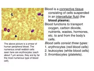
Hematopoiesis • The generation of blood cells takes place in the hematopoietic (from the Greek haima for “blood” and poiesis for “making”) system. • The hematopoietic system encompasses all of the blood cells and their precursors, the bone marrow where blood cells have their origin, and the lymphoid tissues where some blood cells circulate as they develop and mature. • Blood consists of blood cells (i.e., white blood cells, thrombocytes or platelets, and red blood cells) and the plasma in which the cells are suspended.
Leukocytes (White Blood Cells) • The leukocytes, or white blood cells, constitute only 1% of the total blood volume. • They originate in the bone marrow and circulate throughout the lymphoid tissues of the body. • There they function in the inflammatory and immune processes. • They include: • the granulocytes • Neutrophils – 55-65% • Eosinophils – 1-4% • Basophils – 0-1% • the lymphocytes – 20-40% • the monocytes – 3-8%
Hematopoiesis • The blood-forming population of bone marrow is made up of three types of cells: • Self-renewing stem cells • Differentiated progenitor (parent) cells • Functional mature blood cells • All of the blood cell precursors, of the erythrocyte (i.e., red cell), myelocyte (i.e., granulocyte or monocyte), lymphocyte (i.e., T lymphocyte and B lymphocyte), and megakaryocyte (i.e., platelet) series are derived from a small population of primitive cells called the pluripotent stem cells. • A committed stem cell that forms a specific type of blood cell is called a colony-forming unit (CFU). • Under normal conditions, the numbers and total mass for each type of circulating blood cell remain relatively constant. • The blood cells are produced in different numbers according to needs and regulatory factors.
Hematopoiesis • This regulation of blood cells is controlled by a group of short-acting soluble mediators, called cytokines, that stimulate the proliferation, differentiation, and functional activation of the various blood cell precursors in bone marrow. • The cytokines that stimulate hematopoiesis are called colony-stimulating factors (CSFs), based on their ability to promote the growth of the hematopoietic cell colonies from bone marrow precursors. • Lineage-specific CSFs that act on committed progenitor cells include: • erythropoietin (EPO) • granulocyte-monocyte colony-stimulating factor (GM-CSF) • thrombopoietin (TPO) • The major sources of the CSFs are lymphocytes and stromal cells of the bone marrow. • Other cytokines, such as the interleukins, support the development of lymphocytes and act synergistically to aid the functions of the CSFs.
The lymphoid tissues • The lymphoid tissues represent the structures where lymphocytes originate, mature, and interact with antigens. • Lymphoid tissues can be classified into two groups: • The central or generative organs: • Bone marrow, where all lymphocytes arise, and the thymus, where T cells mature and reach a stage of functional competence. • Thymus is also the site where self-reactive T cells are eliminated. • Peripheral lymphoid organs • These are the sites where mature lymphocytes respond to foreign antigens. • They include: • The lymph nodes • The spleen • Mucosa-associated lymphoid tissues • The cutaneous immune system • In addition, poorly defined aggregates of lymphocytes are found in connective tissues and virtually all organs of the body.
Disorders of white blood cells • The number of leukocytes, or white blood cells, in the peripheral circulation normally ranges from 4000 to 10,000/μL of blood. • The term leukopenia describes an absolute decrease in white blood cell numbers. The disorder may affect any of the specific types of white blood cells: • Neutropenia • Lymphopenia • The term leukocytosis describes an absolute increase in in white blood cell numbers and also may affect any of the specific types of white blood cells: • Neutrophilia • Eosiniphilia • Basophilia • Lymphocitosis • Monocitosis
Neutropenia • Neutropenia refers specifically to a decrease in neutrophils. It commonly is defined as a circulating neutrophil count of less than 1500 cells/μL. • Agranulocytosis, which denotes a severe neutropenia, is characterized by a circulating neutrophil count of less than 200 cells/μL. • Neutropenia can be: • Acquired • Congenital • Kostmann’s syndrome • It occurs sporadically as an autosomal recessive disorder, causes severe neutropenia while preserving the erythroid and megakaryocyte cell lineages that result in red blood cell and platelet production. • The total white blood cell count may be within normal limits, but the neutrophil count is less than 200/μL. Monocyte and eosinophil levels may be elevated (compensatory).
Acquired neutropenia • Accelerated removal - removal of neutrophils from the circulation exceeds production • Inflammation • Infection, viral or bacterial • Increased destruction: • Drug-induced granulocytopenia • Treatment of cancer – chemotherapy (e.g., alkylating agents, antimetabolites) • Irradiation • Autoimmune disorders or drug reactions • May cause increased and premature destruction of neutrophils • Splenomegaly • Neutrophils may be trapped in the spleen along with other blood cells • Felty’s syndrome • A variant of rheumatoid arthritis, there is increased destruction of neutrophils in the spleen • Neoplasms involving bone marrow (e.g., leukemias and lymphomas, myeloma)
Acquired neutropenia • Alcoholism • Carentiale states: • Folic acid • Vitamin B12 • Iron • Cooper • Aplastic anemia • All of the myeloid stem cells are affected, resulting in anemia, thrombocytopenia, and agranulocytosis; • Idiopathic neutropenia that occurs in the absence of other disease or provoking influence.
Neutrophilia • Physiological: • In newborns • Pregnancy • In labor • Post-partum • After exercise • Drugs or toxics: • Administration of corticosteroids - may increase the release of neutrophils from the bone marrow and reduce their migration into tissues; • Acute poisoning with Pb, Hg, some venoms; • Reactive neutrophilia - the result of increased release of neutrophils from MB to compensate their high affinity for tissues. It is frequently accompanied by deviation to the left of the leukocyte formula (leukemoid reaction); • Metabolic and endocrine diseases: • Diabetic ketoacidosis • Acute renal failure • Acute gout crise • In some malignant hematologic diseases: CGL, MPCD (PV, ET, CMML)
Eosinophilia • Allergic diseases: asthma, allergic rhinitis, eczema, atopic dermatitis • Parasitic infections • Fungal and other infections • Tuberculosis • Hematologic malignancies (CGL, AL with Eo, LAM2 and 4, rarely in MDS) and nonhematologic (lung, vaginal, skin, stomach carcinoma, malignant melanoma) • Idiopathic - is diagnosis of exclusion • Drugs: aspirin, beta blockers, penicillin, cephalosporins, NSAIDs, etc. • Bacterial infections usually does not cause eosinophilia, but eosinopenia.
Lymphocytosis • Causes of absolute lymphocytosis include: • Acute viral infections, such as infectious mononucleosis (glandular fever), hepatitis and cytomegalovirus infection • Other acute infections such as pertussis • Protozoal infections, such as toxoplasmosis • Chronic intracellular bacterial infections such as tuberculosis or brucellosis • Chronic lymphocytic leukemia • Causes of relative lymphocytosis include: • Age less than 2 years • Acute viral infections • Connective tissue diseases • Thyrotoxicosis • Splenomegaly with splenic sequestration of granulocytes • Exercise • Stress
Infectious mononucleosis • Infectious mononucleosis is caused by the Epstein-Barr virus, a DNA herpes-type virus that infects B lymphocytes. • Patients present with mild to severe adenopathy, hepatosplenomegaly, fever, malaise, pharyngitis, and a characteristic peripheral blood smear demonstrating reactive lymphocytes.
Monocytosis • Monocytosis often occurs during chronic inflammation. Diseases that produce this state: • Infections: tuberculosis, brucellosis, listeriosis, subacute bacterial endocarditis, syphilis, and other viral infections and many protozoal and rickettsial infections; • Blood and immune causes: chronic neutropenia and myeloproliferative disorders; • Autoimmune diseases and vasculitis: systemic lupus erythematosus, rheumatoid arthritis and inflammatory bowel disease; • Malignancies: Hodgkin's disease and certain leukaemias, such as chronic myelomonocytic leukaemia (CMML) and monocytic leukemia; • Recovery phase of neutropenia or an acute infection;
Monocytopenia • The causes of monocytopenia include: • Acute infections • Stress • Aplastic anemia • Hairy cell leukemia • Acute myeloid leukemia • Treatment with myelotoxic drugs • Treatment with glucocorticoids
Surgical, procedure complication Splenectomy Infectious disorders (specific agent) Herpes Zoster Influenza Chickenpox/herpes zoster virus Smallpox (variola) Infected organ, abscesses Inflammatory disorders Neoplastic disorders: Acute myeloid leukemia Hodgkin's disease Myelodysplastic syndrome Chronic myeloid leukemia Polycythemia vera Lymphoma/malignant, non-Hodgkins Allergic, collagen autoimmune disorders Congenital, developmental disorders congenital hemolytic anemia Hereditary, familial, genetic disorders Spherocytosis Vegetative, autonomic, endocrine disorders Hypothyroidism (myxedema) Leukemoid reaction Drugs Foreign protein injection Basophilia
The neoplastic disorders • The neoplastic disorders of hematopoietic and lymphoid origin represent the most important of the white blood cell disorders. • They include three somewhat overlapping categories: • The lymphomas (Hodgkin’s disease and non-Hodgkin’s lymphoma) • The leukemias • Plasma cell dyscrasias (multiple myeloma)
Leukemias • Leukemias are malignant neoplasms of the hematopoietic stem cells with diffuse replacement of bone marrow. • Leukemias are classified according to cell type • Lymphocytic • Myelogenous • and whether the disease is acute or chronic. • The lymphocytic leukemias involve immature lymphocytes and their progenitors that originate in the bone marrow but infiltrate the spleen, lymph nodes, CNS, and other tissues. • The myelogenous leukemias involve the pluripotent myeloid stem cells in bone marrow and interfere with the maturation of all blood cells, including the granulocytes, erythrocytes, and thrombocytes. • The acute leukemias (i.e., ALL, which primarily affects children, and AML, which primarily affects adults) have a sudden and stormy onset, with symptoms of depressed bone marrow function: • Anemia • Fatigue • Bleeding • Infections • Bone pain • Lymphadenopathy • Splenomegaly • Hepatomegaly
Leukemias • The chronic leukemias, which largely affect adults, have a more insidious onset. • CLL often has the most favorable clinical course, with many persons living long enough to die of unrelated causes. • The course of CML is slow and progressive, with transformation to a course resembling that of AML.
Multiple Myeloma • Multiple myeloma is a plasma cell cancer of the osseous tissue and accounts for 10% to 15% of all hematologic malignancies. • It is characterized by the uncontrolled proliferation of an abnormal clone of plasma cells, which secrete primarily IgG or IgA. • There is an atypical proliferation of one of the immunoglobulins, called the M protein, a monoclonal antibody. • Levels of normal immunoglobulins are usually depressed. This contributes a general susceptibility to bacterial infections. • In some forms of multiple myeloma, the plasma cells produce only Bence Jones proteins, abnormal proteins that consist of the light chains of the immunoglobulin molecule. • Because of their low molecular weight, Bence Jones proteins are partially excreted in the urine. • Many of these abnormal proteins are directly toxic to renal tubular structures, which may lead to tubular destruction and, eventually, to renal failure.
Pathogenesis • The cause of multiple myeloma is unknown. • It does not appear to be caused by previous exposure to toxic agents (e.g., solvents such as benzene, paints, pesticides). • Interestingly, an association with human herpesvirus 8 has been described, but the role of this virus in the pathogenesis of the disease remains to be established. • Cytokines are important in the pathogenesis of the disorder. • The multiple myeloma plasma cell has a surface-membrane receptor for interleukin-6, which is known to be a growth factor for the disorder. • Another important growth factor for the myeloma cell is interleukin-1, which has important osteoclastic activity.