Download
1 / 162
1.67k likes | 2.1k Vues
Diseases of White Blood Cells(3). Lecturer :Yiran Ni, MD Department of Pathology China Three Gorges University Email:nyr1986@gmail.com. Dec.2012. What we’ve learned. Leukopenia; Reactive leukocytosis; Lymphadenitis;

E N D
Diseases of White Blood Cells(3) Lecturer :Yiran Ni, MD Department of Pathology China Three Gorges University Email:nyr1986@gmail.com Dec.2012
What we’ve learned • Leukopenia; • Reactive leukocytosis; • Lymphadenitis; • General aspects and classification of neoplastic proliferation of white cell; • Acute myeloid leukaemia
What we’ve learned • Myelodysplastic Syndromes • chronic myeloid leukaemia Ph chromosome • histiocytoses • splenomegaly
Target of this class • Lymphoid neoplasms
Definition Lymphoid neoplasms encompass a diverse group of entities. In many but not all instances, the phenotype of the neoplastic cell closely resembles that of a particular stage of normal lymphocyte differentiation, a feature that is used in the diagnosis and classification of these disorders.
Definition One of the confusing aspects of the lymphoid neoplasms concerns the use of the descriptive terms "lymphocytic leukemia and lymphoma. Leukemia is used for lymphoid neoplasms presenting with widespread involvement of the bone marrow, usually accompanied by the presence of large numbers of tumor cells in the peripheral blood. Lymphoma, on the other hand, is used to describe proliferations arising as discrete tissue masses. Traditionally, these terms were attached to what were felt to be distinct entities. However, the line between the "lymphocytic leukemias" and the "lymphomas" often blurs. Many types of "lymphoma" occasionally present with a leukemic peripheral blood picture accompanied by extensive marrow involvement, and evolution to "leukemia" is not unusual during progression of incurable "lymphomas." Conversely, tumors identical to "leukemias" sometimes arise as soft tissue masses without evidence of bone marrow disease. Hence, when applied to particular neoplasms, the terms "leukemia" and "lymphoma" merely describe the usual tissue distribution of the disease at the time of clinical presentation.
Definition Lymphoma, on the other hand, is used to describe proliferations arising as discrete tissue masses. Traditionally, these terms were attached to what were felt to be distinct entities. However, the line between the "lymphocytic leukemias" and the "lymphomas" often blurs. Many types of "lymphoma" occasionally present with a leukemic peripheral blood picture accompanied by extensive marrow involvement, and evolution to "leukemia" is not unusual during progression of incurable "lymphomas." Conversely, tumors identical to "leukemias" sometimes arise as soft tissue masses without evidence of bone marrow disease. Hence, when applied to particular neoplasms, the terms "leukemia" and "lymphoma" merely describe the usual tissue distribution of the disease at the time of clinical presentation.
Definition Many types of "lymphoma" occasionally present with a leukemic peripheral blood picture accompanied by extensive marrow involvement, and evolution to "leukemia" is not unusual during progression of incurable "lymphomas."
Definition Conversely, tumors identical to "leukemias" sometimes arise as soft tissue masses without evidence of bone marrow disease. Hence, when applied to particular neoplasms, the terms "leukemia" and "lymphoma" merely describe the usual tissue distribution of the disease at the time of clinical presentation.
Lymphoma Lymphoma is a neoplasm of lymphocytes, including T and B cells. Primary sites of lymphoma may be lymphoid tissue or any organ of the body.
The WHO Classification of the Lymphoid Neoplasms • Precursor B-cell neoplasms (immature B cells) • Peripheral B-cell neoplasms (mature B cells) • Precursor T-cell neoplasms (immature T cells) • Peripheral T-cell and NK-cell neoplasms (mature T cells and natural killer cells) • Hodgkin lymphoma (Reed-Sternberg cells and variants).
Classifications • Within the broad group of lymphomas, Hodgkin lymphomais segregated from all other forms, which constitute the non-Hodgkin lymphomas (NHL). • As will be seen, Hodgkin lymphoma is clinically and histologically distinct from the NHLs. • In addition, it is treated in a unique fashion, making the differentiation of Hodgkin lymphoma and NHL clinically important.
Classifications • The other important category of lymphoid neoplasms encompasses the plasma-cell neoplasms, tumors composed of terminally differentiated B cells. • Such tumors most commonly arise in the bone marrow, only rarely involving lymph nodes or producing a leukemic peripheral blood picture. • In addition, as will be seen, much of their patho-physiology is related to the secretion of whole antibodies or immunoglobulin fragments by the tumor cells.
Classifications • The other important category of lymphoid neoplasms encompasses the plasma-cell neoplasms, tumors composed of terminally differentiated B cells. • Such tumors most commonly arise in the bone marrow, only rarely involving lymph nodes or producing a leukemic peripheral blood picture. • In addition, as will be seen, much of their patho-physiology is related to the secretion of whole antibodies or immunoglobulin fragments by the tumor cells.
Classifications • The clinical presentation of the various lymphoid neoplasms is dictated by the anatomic distribution of disease. • Two-thirds of NHLs and virtually all cases of Hodgkin lymphoma present with nontender nodal enlargement (often greater than 2 cm) that can be localized or generalized. • The remaining one-third of NHLs arise at extranodal sites (e.g., skin, stomach, or brain).
Classifications • In contrast, the leukemic forms (lymphocytic leukemia) most commonly come to clinical attention owing to signs and symptoms related to suppression of normal hematopoiesis by tumor cells in the bone marrow. • Lymphocytic leukemias also characteristically infiltrate and enlarge the spleen and liver. • Finally, plasma cell neoplasms involving the skeleton cause local bony destruction and hence often present with pain due to pathologic fractures.
Our focus will be on the subset of neoplasms listed below • Precursor B- and T-cell lymphoblastic leukemia/lymphoma; • Small lymphocytic lymphoma/chronic lymphocytic leukemia; • Follicular lymphomaMantle cell lymphoma • Diffuse large B-cell lymphomas; • Burkitt lymphoma; • Multiple myeloma and related plasma cell dyscrasias • Hodgkin lymphoma.
Principles(1) • Before we discuss the specific entities described in the WHO classification, some important principles relevant to the lymphoid neoplasms need to be emphasized. • 1. Lymphoid neoplasia can be suspected from the clinical features, but histologic examination of lymph nodes or other involved tissues is required for diagnosis.
Principles(2) • 2. Antigen receptor genes rearrange during normal B- and T-cell differentiation through a mechanism that ensures that each developing lymphocyte makes a single, unique antigen receptor. • In most lymphoid neoplasms, antigen receptor gene rearrangementprecedes transformation; hence, the daughter cells derived from the malignant progenitor share the same antigen receptor gene configuration and sequence and synthesize identical antigen receptor proteins.
Principles(2) • In contrast, normal immune responses are polyclonal and thus comprise populations of lymphocytes expressing many different antigen receptors. As a result, analyses of antigen receptor genes and their protein products can be used to distinguish reactive and malignant lymphoid proliferations. In addition, each antigen receptor gene rearrangement produces a unique DNA sequence that constitutes a highly specific clonal marker that can be used to detect small numbers of residual malignant cells after therapy.
Principles(3) • 3. The vast majority of lymphoid neoplasms (80% to 85%) are of B-cell origin, most of the remainder being T-cell tumors; only rarely are tumors of NK origin encountered. • Most lymphoid neoplasms resemble some recognizable stage of B- or T-cell differentiation, a feature that is used in their classification.
Principles(4) • 4. As tumors of the immune system, lymphoid neoplasms often disrupt normal architecture and function of the immune system, leading to immune abnormalities. • Both a loss of vigilance (as evidenced by susceptibility to infection) and breakdown of tolerance (manifested by autoimmunity) can be seen, sometimes in the same patient.
Principles(4) • In a further, ironic twist, patients with inherited or acquired immunodeficiency are themselves at high risk of developing certain lymphoid neoplasms, particularly those caused by oncogenic viruses (e.g., EBV).
Principles(5) • 5. Neoplastic B and T cells tend to recapitulate the behavior of their normal counterparts. • Like normal lymphocytes, transformed B and T cells tend to home to particular tissue sites, leading to characteristic patterns of involvement. • For example, follicular lymphomas proliferate in the B-cell areas of the lymph node, producing a nodular or follicular pattern of growth, whereas T-cell lymphomas typically grow in paracortical T-cell zones.
Principles(5) • As is true of their normal counterparts, lymph node homing of neoplastic lymphocytes is likely regulated by expression of particular chemokine receptors. • Variable numbers of neoplastic B and T lymphoid cells also recirculate periodically through the lymphatics and peripheral blood to distant sites.
Principles(5) • Sensitive molecular techniques have shown that most lymphoid tumors are widely disseminated at the time of diagnosis. The most notable exception to this rule is Hodgkin lymphoma, which is sometimes restricted to one group of lymph nodes.
Principles(6) • 6. Hodgkin lymphoma spreads in an orderly fashion, and as a result staging is of importance in determining therapy. • In contrast, the spread of NHL is less predictable, and as was noted above, most patients are assumed to have systemic disease at the time of diagnosis. Hence, staging in particular NHLs provides useful prognostic information but is generally not as important in guiding therapy as is the case in Hodgkin lymphoma.
Precursor B- and T-Cell Neoplasms (Acute Lymphoblastic Leukemia/Lymphoma )
General aspects • Acute lymphoblastic leukemia/lymphoma (ALL) encompasses a group of neoplasms composed of immature, precursor B (pre-B) or T (pre-T) lymphocytes referred to as lymphoblasts. • The majority (∼85%) of ALLs are precursor B-cell tumors that typically manifest as childhood acute "leukemias" with extensive bone marrow and variable peripheral blood involvement.
General aspects • Malignant pre-B and pre-T lymphoblasts are also morphologically indistinguishable, and subclassification of ALL is thus dependent on immunophenotyping. • Because of their morphologic and clinical similarities, the various forms of ALL will be considered here together.
General aspects • Malignant pre-B and pre-T lymphoblasts are also morphologically indistinguishable, and subclassification of ALL is thus dependent on immunophenotyping. • Because of their morphologic and clinical similarities, the various forms of ALL will be considered here together.
General aspects • Approximately 2500 new cases of ALL are diagnosed each year in the United States, most cases occurring in individuals younger than 15 years of age. • ALL is almost twice as common in whites as in nonwhites and is slightly more frequent in boys than in girls. • than in children.
General aspects • The incidence of pre-B ALL is highest at about the age of 4, perhaps because the number of normal bone marrow pre-B lymphoblasts (the cell of origin) peaks in early childhood. • Similarly, the peak incidence of pre-T ALL is in adolescence, the age when the thymus reaches its maximal size. • Both pre-B and pre-T ALL occur in adults of all ages, but much less frequently than in children.
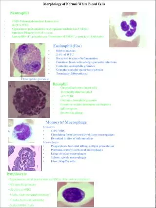
Morphology • Because of different responses to chemotherapeutic agents, it is of great practical importance to distinguish ALL from acute myelogenous leukemia (AML), a neoplasm of immature myeloid cells that may cause identical signs and symptoms. • Compared to myeloblasts, lymphoblasts have condensed chromatin, inconspicuous nucleoli, and scant agranular cytoplasm.
lymphoblasts have condensed chromatin, inconspicuous nucleoli, and scant agranular cytoplasm
Morphology • However, these morphologic distinctions are not absolute, and definitive diagnosis relies on detection of B and T lymphocyte-specific markers with antibodies. • Histochemical stains can also be helpful, as lymphoblasts (in contrast to myeloblasts) lack peroxidase-positive granules and often contain cytoplasmic aggregates of periodic acid-Schiff (PAS)-positive material.
Morphology • As has been noted, ALLs with lymphomatous presentations are mostly of pre-T cell type. • Many pre-T ALLs (50% to 70%) are associated with mediastinal masses stemming from thymic involvement, and lymphadenopathy and splenomegaly are also more prevalent in this subtype.
Morphology • Regardless of phenotype, the histologic appearance of ALL is similar. • Normal tissue architecture is completely effaced by lymphoblasts having scant cytoplasm anda nuclei somewhat larger than those of small lymphocytes. • The nuclear chromatin is delicate and finely stippled, and nucleoli are either absent or inconspicuous.
Morphology • In many cases, the nuclear membrane shows deep subdivision, imparting a convoluted (lobulated) appearance. • In keeping with its aggressive growth, the tumor shows a high rate of mitosis, and as with other tumors having a high mitotic rate (e.g., Burkitt lymphomas), a "starry sky" pattern can be produced by interspersed benign tingible body macrophages that have ingested the debris of dying neoplastic cells.
Immunophenotype • Immunostaining for terminal deoxynucleotidyltransferase (TdT), a specialized DNA polymerase that is expressed only by pre-B and pre-T lymphoblasts, is positive in >95% of cases
Immunophenotype • Precursor B ALL cells are arrested at stages preceding surface expression of Ig. • The leukemic blasts almost always express the pan B-cell molecules CD19 and CD10. • In very early pre-B cell ALL, CD19 is the only B cell-specific marker present. • Early pre-B ALL is distinguished from late pre-B ALL by the absence of cytoplasmic IgM heavy chain (μ chain) in the former.
Immunophenotype • Precursor T ALL cells are arrested at early stages of T-cell development. • In most cases, the cells are CD1+, CD2+, CD5+, and CD7+. • Early pre-T cell tumors are usually negative for surface CD3, CD4, and CD8, whereas late pre-T cell tumors are positive for these markers.
Cytogenetics and Molecular Genetics • Approximately 90% of patients with ALL have numerical or structural changes in the chromosomes of the leukemic cells. • Most common is hyperploidy (>50 chromosomes), but also polyploidy, and t(12;21), t(9;22) (Philadelphia chromosome) and t(4;11) translocations.
Clinical Features • It should be emphasized that although ALL and AML are immunophenotypically and genotypically distinct, they usually present with very similar clinical features. • In both diseases, an accumulation of neoplastic "blast" cells in the bone marrow suppresses normal hematopoiesis by physical crowding, competition for growth factors, and other poorly understood mechanisms. • .
Clinical Features • This results in anemia, neutropenia, and thrombocytopenia, which underlie the major clinical features of both ALL and AML. • These common features and those more characteristic of ALL are listed below: • 1. Abrupt stormy onset: Patients present within days to a few weeks of the onset of symptoms.