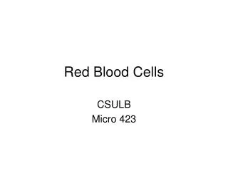
RED BLOOD CELLS
RED BLOOD CELLS. by Mary Yvonnette C. Nerves, MD, FPSP. Erythropoiesis. A process by which early erythroid precursor cells differentiate to become the mature RBCs Primary regulator: ERYTHROPOIETIN - stimulates red cell precursors at all levels of maturation to hasten the maturation process


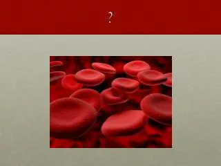
RED BLOOD CELLS
E N D
Presentation Transcript
RED BLOOD CELLS by Mary Yvonnette C. Nerves, MD, FPSP
Erythropoiesis • A process by which early erythroid precursor cells differentiate to become the mature RBCs • Primary regulator:ERYTHROPOIETIN - stimulates red cell precursors at all levels of maturation to hasten the maturation process - responsible for stimulating the premature release of reticulocytes into the bloodstream.
Erythropoiesis • Total erythropoiesis: - total number of red blood cells (RBCs) - measured by the myeloid-erythroid (M:E) ratio from aspirate smears plus the estimate of cellularity from biopsy sections
Effective erythropoiesis: - number of viable and functional RBCs available for physiologic needs - reflects the balance between the number of cells produced and their life span - measured by the reticulocyte count, which is normally 1% of the total RBC count
Stages of Maturation • Pronormoblast (Rubriblast) • Basophilic Normoblast (Prorubriblast) • Polychromatophilic Normoblast (Rubricyte) • Orthochromatic Normoblast (Metarubricyte) • Reticulocyte • Erythrocyte
Pronormoblast • Earliest recognizable and largest cell of the erythrocyte series • Morphology: - Size: 12 – 20 um - Nucleus: large round, oval, dark violet; fine chromatin; 1 – 2 nucleoli - Cytoplasm: deep blue spotty, basophilic w/ a perinuclear halo - N/C Ratio: 8:1 - BM (%): 1
Basophilic Normoblast • Hemoglobin synthesis begins at this stage • Morphology: - Size: 10 – 15 um - Nucleus: large round to sl oval; condensed, coarse chromatin; 0 – 1 nucleoli - Cytoplasm: deeply basophilic; clusters of free ribosomes - N/C Ratio: 6:1 - BM (%): 1-4
Polychromatic Normoblast • Increased production of hemoglobin pigmentation and decreasing amounts of RNA • Last stage in which the cell is capable of mitoses • Morphology: - Size: 10 - 15 um - Nucleus: round nucleus, deep staining, may be centrally or eccentrically located; coarse & clumped chromatin - Nucleoli: 0
Morphology: - Cytoplasm: abundant blue-gray (RNA) to pink-gray (hemoglobin) - N/C Ratio: 4:1 - BM (%): 10-20
Orthochromatic Normoblast • The last nucleated stage • Cannot synthesize DNA and cannot undergo cellular division • The NRBC sometimes seen in the peripheral circulation
Morphology: - Size: 8 - 10 um - Nucleus: small pyknotic nucleus; dense chromatin; 0 nucleoli - Cytoplasm: abundant red-orange cytoplasm uniform in color - N/C Ratio: 1:2 - BM (%): 5-10
Reticulocyte • Slightly larger than the mature RBC with residual amts of RNA • Reticulocyte count: an index of bone marrow activity or effective erythropoiesi • Morphology: - Size: 8 - 10 um - Nucleus: anucleate cell containing small amt of basophilic reticulum (RNA) - Nucleoli: 0 - Cytoplasm: large amt of blue-pink staining hemoglobin cytoplasm
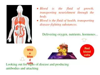
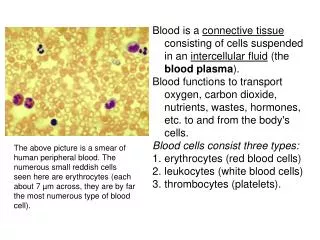
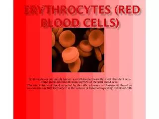
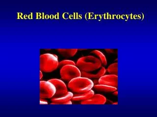
Erythrocyte • A biconcave 6 – 8 um disc • Life span: 120 days • Main function: to transport hemoglobin, a protein that delivers oxygen from the lungs to tissues and cells • Contains 90% hemoglobin and 10% H2O • normal conc of RBCs varies w/ age, sex & geographic distribution
Morphology: - Size: 7 - 8 um - Nucleus: anucleated cell - Nucleoli: 0 - Cytoplasm: pink staining, zone of central pallor is 1/3 of cell diameter devoid of hemoglobin - N/C Ratio: NA
Hemoglobin: Structure & Function • A conjugated protein that serves as the vehicle for the transportation of O2 and CO2 • When fully saturated, each gram of Hgb can hold 1.34 mL of O2 • A molecule of Hgb consists of 2 pairs of polypeptide chains (“globin”) and 4 prosthetic heme grps each contg 1 atom of ferrous iron
DESCRIPTION of TERMS SIZE DESCRIPTORS • Anisocytosis: variation in the sizeof the RBCs due to a pathologic condition
Normocytic: normal sized biconcave disc RBC - normal MCV
Microcytic: Smaller RBCs less than 6 um - MCV < 80 fl - Defect / Change: abn size due to failure of hgb synthesis - Dse: IDA, Thalassemia, Chronic dse • Macrocytic: Larger RBCs greater than 9um - MCV > 90 fl - Defect / Change: impaired DNA synthesis / stress erythropoiesis - Dse: Megaloblastic anemia / liver dse / MDS / Alcoholism / Malaria
Macrocytic Microcytic
CHROMICITY DESCRIPTORS • Normochromic: normal in color; pale central area occupies less than 1/3 - Defect / Change: normal amt of Hgb - Normal indices • Hypochromic: an RBC that has a decreased Hgb complement - central pallor exceeds 1/3 of diameter of cell - Defect / Change: reduced Hgb content ( MCHC) - Assoc conditions: IDA / Thalassemia
“Hyperchromic”: no central pallor - Defect / Change: greater than normal MCHC - Assoc condition: Spherocytosis “Hyperchromic” Hypochromic
Polychromasia: blue-gray coloration - Defect / Change: presence of RNA - Assoc condition: increased erythropoietic activity / hemorrhage / hemolysis
SHAPE DESCRIPTORS • Poikilocytosis:variation in shape of the RBC - Defect / Change: irreversible alteration of membrane - Assoc conditions: Anemia / Hemolytic states
Discocyte: normal biconcave erythrocyte - 6 – 8 um diameter; 0 – 2 um thickness - Aka: Normocyte Normal Red Cells (SEM)
Acanthocyte: spheroid w/ 3 – 12 irreg spikes or spicules - Aka: spur cell - decreased cell volume - Defect / Change: inc ratio of chole to lecithin - Assoc conditions: end-stage liver dse Pyruvate kinase def Hemolytic anemia Abetalipoproteinemia
Blister cell: contains 1 or more vacuoles - Aka: Bite cells - thinned periphery - Defect / Change: formed by removal of Heinz bodies - Assoc conditions: Hemolytic episodes G6PD def Hemoglobinopathies
Codocyte : peripheral rim of Hgb surr by clear area & central hemoglobinized area (bull’s eye) - Aka: target cell - Defect / Change: excess of surface to volume ratio - Assoc conditions: Hemoglobinopathies Thalassemia Liver dse Postsplenectomy
Dacryocyte: teardrop or pear-shaped w/ single elongated point or tail - Aka: tear drop cell - Defect / Change: squeezing & fragmentation during splenic passage - Assoc conditions: Myeloid metaplasia Thalassemia Megaloblastic anemia Hypersplenism
Drepanocyte: crescent-shaped cell that lacks zone of central pallor - Aka: Sickle cell - Defect / Change: polymerization of deoxygenated Hgb - Assoc conditions: Sickle cell anemia SC disease S-thalassemia