Download
1 / 63
850 likes | 2.41k Vues
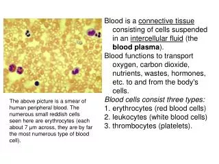
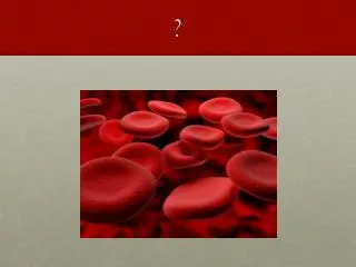
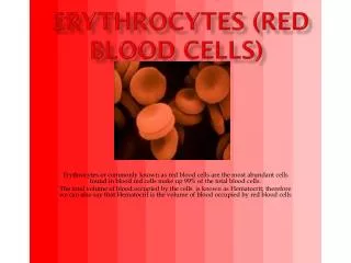
Red Blood Cells (Erythrocytes). Er ythrocytes (RBC). Structure Biconcave disc shape Includes Hemoglobin Lipid s , ATP, carbonic anhydrase Function O 2 and CO 2 transport. Er ythrocytes (RBC). Most abundant type of blood cells 4- 6 million /mm 3 anemi a ; number of RBC below normal

E N D
Erythrocytes (RBC) • Structure • Biconcave disc shape • Includes • Hemoglobin • Lipids, ATP, carbonic anhydrase • Function • O2 and CO2 transport
Erythrocytes (RBC) • Most abundant type of blood cells • 4-6million/mm3 • anemia; number of RBC below normal • polycythemia (erythrocytosis); number of RBC above normal range • No nucleus, mitochondria and ribosom • No protein synthesis • Energy is obtained through glycolisis, but there is no glycogen storage • Normal diameter 7-8 , volume 78-94 3 • normocyte - microcyte - macrocyte
Energy use and source in RBC • Energy is used mainly for two functions: • Continuation of membrane shape - active transport • Homeostasis of intracellular oxidation-reduction • Only source of energy is glycolysis
RBC Membrane • The same as other biological membranes • Cannot synthesize its own cell membrane • Actin and spectrin provide strength • Contractility in the presence of Ca+2 ions • Changes in viscosity • Na+-K+ATPaseand Ca+2ATPase activity • Carbohydrates on the membrane are to do with blood types (antigens)
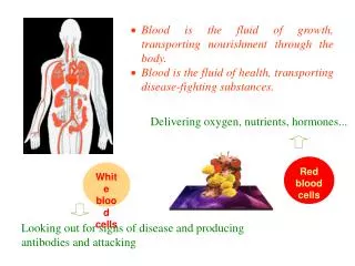
Hemoglobin (Hb) • Red color protein that carries oxygen • 1 g Hb binds 1,3 mlO2; 14,8 g Hb binds 20 ml O2 • Normal range of hemoglobin: 14-16 g/dl • In adults, about 25-30 trillion RBC, and 900 g Hb • Hb constitutes 1/3 of erythrocyte weight
Hemoglobin • 4 globin molecules:carries CO2 (role of carbonic anhydrase) • 4 heme molecules:carries O2 • Iron is required for oxygen transportation
Hematopoiesis (Hemopoiesis) • Production of blood cells • Stem cells: All formed elements of blood stem from a colony of cells • Proerythroblast: forms erythrocytes • Myeloblast: forms neutrophils, eosinophils and basophils • Lenfoblast: forms lymphocytes • Monoblast: forms monocytes • Megakaryoblast: forms thrombocytes
Embryonic Stem Cells Human ESC
What are stem cells? • They are of many types: epidermal, intestinal, hematopoietic, etc. • The defining properties of a stem cell are: • It is not terminally differentiated. • It can divide without limit. • When it divides, the daughter cell has a “choice”: • Remain a stem cell, or • Terminally differentiate.
Potentiality of Stem Cells “Stem cells” have varying potentials: • Totipotent cells.Fertilized oocyte (zygote) & progeny of the first two cell divisions. Cells able to form the embryo and the trophoblast of the placenta. • Pluripotent cells. After about 4 days, the blastocyst forms; embryonic stem cells obtained from the inner cell mass, which becomes the embryo, are pluripotent, able to differentiate into almost all cells of the three germ layers – but not into an embryo. • Multipotential cells. Found in most tissues, these cells can produce a limited range of differentiated cell lineages appropriate to their location. (Hematopoietic stem cells from the bone marrow exemplify multipotential cells.) • Unipotential cells. Cells capable of generating only one cell type (epidermal stem cells, adult liver stem cells).
RBC Formation (Erythropoiesis) • In the adult, all blood cell formation (including erythropoiesis) occurs in the red bone marrow • All blood cells develop from stem cells called hemocytoblasts Red Blood Cells Monocytes Platelets Granulocytes Myeloid Stem Cell Hemocytoblast Lymphoid Stem Cell Lymphocytes
Formation of multiple different blood cells from pluripotent hematopoietic stem cell (P-HSC)
Erythropoiesis (production of RBC) Weeks 16-20 Week 2 Week 6 Birth Week 8 Myeloid phase Spleen Hepatic phase Mesoblastic phase Myeloid phase Prenatal Life Postnatal Life
Prenatal period-1 • Mesoblastic phase • It starts in the vitellus sac at the second week • These are erythrocytes with nuclei • Embriyonic hemoglobins: • Hb Gower I, Gower II and Portland 1
Prenatal period-2 • Hepatic phase • Liver takes part starting from the week 6 • Spleen is involved starting from the week 8 • Fetal hemoglobin: • HbF (2 and 2 )
Prenatal period-3 • myeloid phase • Starts between the weeks 16 and 20 in the bone marrow • All bones • Hemopoiesis • HbA (2 and 2 ): mature hemoglobin
Postnatal Period • Only in the bone marrow(myeloid) • All bones contribute up to the age of 5 • Later, erythropoiesis regresses from the distal to the proxymale • vertebra, sternum, costa, cranium and femur • Red bone marrow turns into yellow marrow (reversible) • Lipid infiltration
Erythropoiesis • BFU-E and CFU-E cells • Response to different levels of EPO • proerythroblast • basophil erythroblast • Polychromatophil erythroblast • Orthochromatophil erythroblast • Reticulocyte • Erythrocytes
Regulation of Erythropoiesis • Primary stimulus is hypoxia • Tissue oxygenation • Blood flow • Blood hemoglobin levels • Oxygen saturation of hemoglobin • Affinity of hemoglobin to oxygen
Erythropoietin (EPO) • Fetus and newborn • 85% Liver • hepatocyte, Kupffer cells, endothelial cells • 15% kidney • Endothelial cells, glomerulus, JGH, proxymal tubule • Adult • 85% kidney • 15% liver and other tissues • glomus caroticum, macrophages
Erythropoiesis • Erythropoietin: a hormone that stimulates erythropoiesis • It stimulates both differentiation and maturation
Maturation of red blood cells – Vit B12 and Folic acid • Both vitamins are necessary for DNA synthesis • Deficiency of either of these vitamins causes maturation failure in the process of erythropoiesis • Pernicious anemia – Megaloblastic anemia • Vit B12 absorption and storage • Intrinsic factor: released from parietal cells in the stomach • Complex of Vit B12 + intrinsic factor cannot be digested by the enzymes in the stomach • Lack of intrinsic factor causes serious absorption abnormalities of Vit B12
Hemoglobin (Hb) • Normal range of hemoglobin: 14-16 g/dl • Men: 16 g/dl = 21 ml O2 /dl blood • Women: 14 g/dl = 19 ml O2 /dl blood
Formation of Hemoglobin • 2 succinyl-CoA + 2 glycine = pyrrole • 4 pyrrole protoporphyrin IX • protoporphyrin IX + Fe++ Heme • Heme + globin hemoglobin chain (alpha or beta) • 2 alpha + 2 beta chains Hemoglobin A
Hemoglobin • Hemoglobin synthesis begins in the proerythroblasts and continues until reticulocytes • It consists of iron containing heme and globin • Hemoglobin A contains 2a ve 2b chain • There are different types of these chains (a, b, d ve g) • Hb molecule binds to oxygen loosely and reversibly
Hemoglobin A • 4 globin molecules:carries CO2 (role of carbonic anhydrase) • 4 hem molecules:carries O2 • Iron is required for oxygen transportation
Lifecycle of an RBC • RBCs are subjected to incredible mechanical stress. • Why are they unable to synthesize replacements for damaged parts? • After ≈120d, the RBC cell membrane ruptures, or the damage is detected by phagocytic cells and the RBC is engulfed. • If the RBC hemolyzes, its contained Hb will be excreted by the kidneys A macrophage phagocytizing multiple RBCs
Life span and destruction of RBCs • Because of lack of nuclei, they cannot divide and grow • They have a life span of 120 days • Old erythrocytes are destructed in the spleen, liver and bone marrow • Hemoglobin is broken down to heme and globin • Iron part of heme is stored for re-use • Porphyrin part of hemoglobin is converted to bilirubin and secreted into bile
Iron – Fe+2,+3 • 50 in men and 35 mg/kg in women (total 4-5 gr) • 60-65% in hemoglobin • 4% myoglobin • 1% bound to plasma transferrin • The rest is in ferritin or hemosiderin • Amount of iron bound to transferrin 110-130 g • Daily need of iron lost through urine, feces and bleeding should be compensated
Transferrin - siderofilin • It is a 1-globulin • It has two sides to bind iron (ferri) • One side leaves iron to the liver and the other to bone marrow for hemoglobin synthesis • Saturation of transferrin with iron (normally 35%)
Absorption of iron • Iron is absorbed in ferro (+2) form • Absorption in all parts of the small intestine • It binds to apoferritin in the intestinal cells and stored as ferritin • Iron in ferritin is in the form of ferri (+3) • Liver is the largest storage site • Homeostasis is maintaned through the intestinal depot
Iron Overload Disease • hemosiderin • hemochromatosis • Bronze diabetes • Cirrhosis • Cancer • Gonadal atrophy
Iron Deficiency • Hepatic diseases • Deficiency of reducing substances in the digestive tract • Ca+2, ascorbic acid, lactic acid, pyruvate, glucose and sorbitol • Excessive Ca+2 • Oxalate, phytate, phosphate • Important symptom: geophagy
Other factors • Time of iron supplementation • Injections in cases of absorption problems • Overload of iron facilitates production of microorganisms • transferrin and lactoferrin are anti-microbial
Anemia • Decrease in RBC count and/or amount of Hb • Reduced capacity of blood to carry O2 • Tachycardia, tachypnea • Tiredness, feeling cold • General causes • Blood loss • Reduced erythropoiesis • Increased destruction of RBC • Inadequate of production of EPO
Classification of Anemias • Hemorrhagic anemias; blood loss • Hemolytic anemias; hemolysis • Vitamin deficiency anemias (megaloblastic anemia) • Iron deficiency anemia • EPO deficiency caused anemia • Aplastic anemia (Fanconi anemia)
Hemolytic Anemias • Intracorpuscular causes • hereditary spherocytosis • sickle cell anemia; HbS • thalassemia (Mediterranian anemia, cooley anemia) • glucose-6-phosphate dehydrogenase deficiency • Pyruvate kinase deficiency • Paroxysmal nocturnal hemoglobinuria • Extracorpuscular causes • Blood transfusion – autoantibodies
Sickle Cell Anemia • Seen in black population • A mutation in the βchain of globin results in HbS • This hemoglobin precipitates as long crystals in RBC when it exposes to O2 • As a result, RBC become sickle shaped and may cause blockage in small blood vessels
Sickle cell anemia (SCA) • In SCA, aa valine takes the place of glutamic acid at the Hemoglobin beta polypeptide chain