Download
1 / 66
750 likes | 1.55k Vues
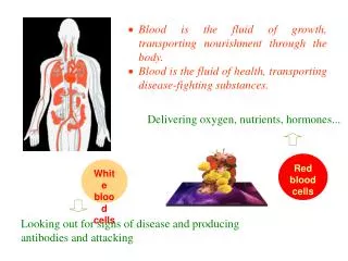
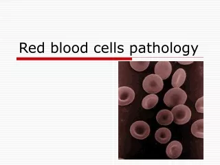
Red Blood Cells, Anemia and Polycythemia. Prof. dr. Zoran Vali ć Department of Physiology University of Split School of Medicine. Red Blood Cells (Erythrocytes). functions: transport of hemoglobin (O 2 ) in some animals it circulates as free protein

E N D
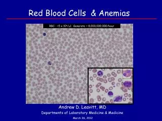
Red Blood Cells, Anemia and Polycythemia Prof. dr. Zoran Valić Department of Physiology University of Split School of Medicine
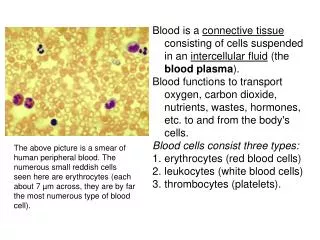
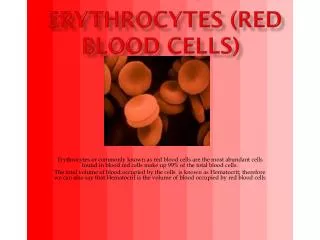
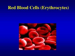
Red Blood Cells (Erythrocytes) • functions: • transport of hemoglobin (O2) • in some animals it circulates as free protein • in humans within RBC – loss by filtration 3% • large quantity of carbonic anhydrase (CO2 and H2O) • an excelent acid-base buffer (proteins)
biconcave discs (φ=7,8 μm; V=90-95 μm3) • shape can change remarkably (squeeze through capillaries, excess of membrane) • M = 5,2x10¹² • F = 4,7x10¹² • chemoglobin in RBC < 340 g/L • Ht = 40-45% • chemohlobin in blood = 160-140 g/L
yolk sac (few early weeks) • liver; spleen and lymph nodes(middle trimester of gestation) • bone marrow • beyond the age of 20 most RBC are produced in membranous bones (vertebrae, sternum, ribs and ilia)
growth inducers – proteins which control growth and reproduction of stem cells • interleukin-3 – promotes growth and reproduction of virtually all stem cells • differentiation inducers (low oxygen, infectious diseases)
1% bone marrow
tissue oxygenation – most essential regulator (viscosity) • hemorrhage, x-ray therapy, high altitudes, cardiac failure, lung diseases • erythropoietin (glycoprotein; 34000) • 90% is formed in kidneys (unknown, liver) • fibroblast-like interstitial cells surrounding the tubules? • renal tissue hypoxia (and some other) HIF-1 erythropoietin • quick secretion (min – 24 h), RBC in 5 days • production of proerythroblasts, speeding up
erythropoietic cells are among the most rapidly growing and reproducing cells • person’s nutritional status • vitamin B12 and folic acid (thymidine) • macrocytes – flimsy membrane and irregular, large shape – shorten life span (1/2-1/3 normal) • B12 – pernicious anemia (atrophic gastric mucosa; parietal cells – intrinsic factor) • folic (pteroylglutaminic) acid – widely spread but destroyed during cooking – sprue
Formation of Hemoglobin • begins in proerythroblasts and continues even into the reticulocyte stage • succinyl-CoA from Krebs metabolic cycle • alpha, beta, gamma and delta chains • most common – hemoglobin A (2 alpha, 2 beta chains) • each hemoglobin molecule transports 4 molecules of oxygen
sickle cell anemia –the amino acid valine is substituted for glutamic acid at one point in each of the two beta chains • 15 μm elongated crystals in low oxygen environment • loosely and reversibly combining with O2 • “coordination bond”, molecular oxygen
Iron Metabolism • hemoglobin, myoglobin, cytochrome-oksidase, peroxidase and catalase • total iron in the body – 4-5g (65% in hemoglobin, 4% in myoglobin, 15-30% in reticuloendothelial system and liver parenchymal cells)
transferrin molecule binds strongly with receptors in the cell membrane s of erythroblasts in bone marrow – endocytosis • inadequate quantities of transferrin – failure to transport iron to the erythroblasts – hypochromic anemia
Absorption of Iron • liver secretes moderate amounts of apotransferrin into the bile – transferrin (with the iron, pinocytosis into enterocyts, plasma transferrin) • absorption is slow and limited; total body iron is regulated mainly by altering the rate of absorption
Life Span of RBC • average circulating time 120 days • cytoplasmic enzymes: • maintaining pliability of the cell membrane • maintain membrane transport of ions • keep the iron in ferrous, rather than ferric form • prevent oxidation of the RBC proteins • many RBC self-destruct in the spleen (when squeezing through the red pulp)
hemoglobin is phagocytized by macrophages (Kupffer cells of the liver) iron and bilirubin (from porphyrin portion)
Anemias (deficiency of hemoglobin) • microcytichypochromic anemia – blood loss anemia (acute and chronic) • aplastic anemia – bone marrow aplasia (high-dose radiation, chemotherapy, drugs, toxic chemicals – insecticides or benzene) • megaloblastic anemia (lack of B12 (pernicious) or folic acid)
hemolytic anemia (abnormalities (hereditary) of RBC) • hereditary spherocytosis (small and spherical RBC) • sickle cell anemia (hemoglobin S, crisis) • erythroblastosis fetalis
Effects of Anemia on Circulation • viscosity of blood depends largely on RBC • fall in blood viscosity decrease in total resistance (added tissue hypoxia – vasodilation) increase in CO (3-4x) increased pumping workload on the heart • problems during exercise – acute cardiac failure
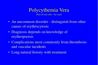
Polycythemia • secondary polycythemia – due to hypoxia (at high altitude, cardiac failure) – 6-7 x 10¹² (30%) • polycythemiavera (erythremia) – 7-8 x 10¹² (Ht = 60-70%) – genetic aberration in the hemocytoblastic cells • increased viscosity – CO almost normal (decreased venous return, but increased blood volume), ruddy complexion with a bluish (cyanotic) tint to the skin)
Antigenicity • first attempts were unsuccessful • transfusion reaction and death • blood posses antigenic and immune properties • at least 30 commonly occurring, and hundreds of other antigens • most of antigens are week, used to establish parentage • systems: O-A-B and Rh
OAB system is discovered by Austrian scientist Karl Landsteiner 1900. (three types, awarded Nobel prize 1930; simultaneously with Czech serologist Jan Janský) • also with Alexander S Wiener identified Rh factor 1937.
O-A-B Blood Types • antigens A i B (also called agglutinogens – cause blood cell agglutination) occur on the surface of the RBC • because of the way of inheritance people may have neither of them on their cells, they may have one or they may have both simultaneously
when neither A or B agglutinogen is present – blood (person) is blood type O • only agglutinogen A – blood is type A • only agglutinogen B – blood is type B • when both agglutinogens are present – blood is type AB
antigen H – essential precursor of OAB blood antigens • located on chromosome 19, posses 3 exons which are coding enzyme fucosyltransferase • enzyme creates H antigen on RBC • carbohydrate chain: β-D-galactose, β -D-N-acetilglucosamine, β -D-galactoseiα-L-fucose (connection with protein or ceramid)
OAB locus is on chromosome 9, has 7 exons • exon 7 is the biggest and contains the greatest portion of coding sequence • OAB locus has 3 allele types: O, A, B
allele A codes glycosyltransferase which bindes N-acetylgalactosamine on D-galactose end of H antigen • allele B codes glycosyltransferase which bindesα -D-galactose on D-galactose end of H antigen • allele 0 has deletion in exon 6 – loss of enzimatic activity – only H antigen is present
Relative Frequencies of the Different Blood Types: 0 47% A 41% B 9% AB 3% • there are 6 different allele types among white population: (A1, A2, B1, O1, O1v i O2), in Asian population B type is more frequent
Agglutinins • antibodies directed at agglutinogens • immediately after birth – not present • they are formed 2-8 month after the birth • maximum titer is reached 8-10 years of age • gamma-globulins (IgMiIgG) • why are they produced? • environmental antigens (bacteria, viruses, plants, foods)
for anti-A agglutinins – influenza • for anti-B agglutinins – gram-negative bacteria (E. coli) • “light in the dark” theory – viruses during replication process incorporate parts of host membrane
Agglutination Process • agglutinins have 2 (IgG) or 10 (IgM) binding sites for agglutinogens • attaching to two or more RBC – bounding together (clump of cells) – agglutination • plugging of small blood vessels throughout the circulation – physical distortion of the cells or phagocytosis – hemolysisof the RBC
Acute Hemolysis • on rare occasion • hemolysis occurs immediately in circulating blood • activation of the complement system – release of proteolytic enzymes (the lytic complex) – rupture of the cell membranes (existence of high titer of IgM antibodies – hemolysins)
Blood Typing • blood typing and blood matching • RBC are separated from the plasma and diluted with saline; mixing with anti-A and anti-B agglutinins