Download
1 / 24
240 likes | 394 Vues
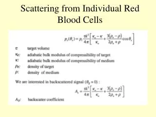
Scattering from Individual Red Blood Cells. Scattering from Ensembles of Red Blood Cells. Scattering from Ensembles of Red Blood Cells. The Doppler Effect. Moving source. Moving receiver. ‘Pulse-echo’: . Continuous wave Doppler System: getting directionality I.

E N D
The Doppler Effect Moving source Moving receiver ‘Pulse-echo’:
Pulsed wave Doppler: Approach Question: what is really being measured here?
Pulsed wave Doppler:Signals Reference oscillator
Measurement limitations: aliasing Well sampled At Nyquist (PRF/2) Aliased
Measurement limitations: velocity resolution limits Velocity resolution Frequency resolution Fres = 1/(total time) = PRF/(N) where N=number of pulses Using the Doppler equation… Vres = c*PRF/(2*Ftrans*N) Increasing N therefore improves resolution, but total time must be less than that of expected changes to flow (e.g. due to cardiac cycle)
Measurement limitations: tranverse motion Fundamental trade-off between localization and velocity resolution
Colour flow: how it works Demodulated Audio (time domain) Audio (freq. domain) For pulsed wave Doppler we had: And calculated full spectra (i.e. velocity distributions in a time window) with: For colour flow, we use the autocorrelator to calculate the mean velocity (Jensen ’96): N=# pulses Is=Iaudio
Demodulated Audio (time domain) Audio (freq. domain) Power Doppler: how it works For power Doppler, we use the audio frequency data to calculate the mean power: N=# pulses Is=Iaudio The power is proportional to the volume of moving blood
Colour flow: ‘clutter’ filtering Prior to power and velocity estimation, a high pass filter (e.g. FIR, IIR, regression) is applied to remove the tissue signals - the ‘clutter’ filter can have a fundamental impact on flow detection
Flow imaging processing overview Velocity Est. Thresholding/ Flow decision Velocity image Raw Data ‘ensemble’ Clutter filter Power Est. Power image Variance Est. Variance image Power est. Bscan - Ensembles generally 4-16 pulses along a beam direction - process repeated across image plane
Colour flow: limitations • - noise and small number of pulses results in vessel detection limits • - ‘flash’ artefacts due to transient tissue/transducer motion • filtering results in increased loss of slower flow • As with pulsed-wave Doppler: • -aliasing • -depth vs velocity limits (more stringent due to 2D image acquisition)