Immune Cell Distribution in Draining Lymph Nodes: Monocytes, B Cells, and T Cell Subtypes
10 likes | 135 Vues
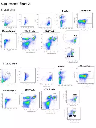
This figure illustrates the composition of immune cell subsets in draining lymph nodes (DLNs), highlighting the roles of monocytes, B cells, macrophages, CD8+ T cells, and CD4+ T cells. Two distinct profiles are presented: (a) showing the presence of mock monocytes alongside various T cell categories, and (b) displaying the distribution of immune cells in DLNs expressing the costimulatory molecule 41BB. The data are represented using immunoelectron microscopy (IEM), electron microscopy (EM), and confocal microscopy (CM) techniques, emphasizing the cellular landscape in immune response.

Immune Cell Distribution in Draining Lymph Nodes: Monocytes, B Cells, and T Cell Subtypes
E N D
Presentation Transcript
Supplemental figure 2. a) DLNs Mock Monocytes B cells Macrophages CD8 T cells CD4 T cells IEM EM CM b) DLNs 41BB Monocytes B cells CD4 T cells CD8 T cells Macrophages IEM EM CM