Facial Palsy
David Kuykendall, MD University of Nevada School of Medicine 2013. Facial Palsy. Caused by Malformation Damage Agenesis Affects the 7 th Cranial Nerve Muscles for Facial Expression Taste of anterior 2/3 of tongue. Congenital Facial Palsies.

Facial Palsy
E N D
Presentation Transcript
David Kuykendall, MD University of Nevada School of Medicine 2013 Facial Palsy
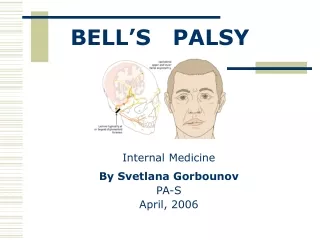
Caused by • Malformation • Damage • Agenesis • Affects the 7th Cranial Nerve • Muscles for Facial Expression • Taste of anterior 2/3 of tongue Congenital Facial Palsies
Often noticed in first 24 hours of life during babies’ first cries • Can present as poor feeding • Evaluate baby for stroke: • Strength of movement, Reflexes, and Sensation of extremities on the affected side. • If normal: suspect congenital facial palsy • Ear tags/pits, malformed external ears, and/or facial asymmetry may indicate Brachial Arch Syndrome Presentation
Family History • Pregnancy Risks: infections, substance use/abuse, trauma, PMHx, pets/environment • Associated symptoms: doesn’t seem to hear/see well, neurological dysfunctions, fever, poor feeding, etc. • Exam: otologic, neurologic, ocular, dermatologic and oral exams History and Physical
Birth Trauma (Most Common) • Brachial Arch Syndromes • Moebius Syndrome • Hemifacial Microsomia • Goldenhar’s Syndrome • Bells Palsy • Tumor Causes
Injuries that occur during birthing process or facial pressure on sacrum from position of baby in the uterus • Risk Factors: LGA, Gestational diabetes, post-term, vaginal deliveries, intrauterine trauma, assisted deliveries (vacuum and forceps) • Usually resolve in first week of life • Few (less than 5%) cases of permanent nerve damage with no facial paralysis improvement (Lundstrom, 2002) Birth Trauma
Development (abridged) • During fourth week of life the embryo develops brachial (pharyngeal) arches • The Second Brachial Arch gives rise to CN VII • During the sixth week of life the external ear develops from the 1st and 2nd Brachial Arch • Malformations are often accompanied by facial nerve dysfunctions as malformation of the ear can result in disrupted growth of CN VII (Backous, 1991) • Causes: • Genetic Mutation • Vascular Disruption • Trauma or Blood Loss during pregnancy Brachial Arch Syndromes
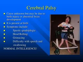
Bilateral congenital facial paralysis • Can’t smile, expressionless face, limited eye movement, feeding difficulties (most need feeding tubes), and high risk of aspiration • Normal Intelligence • Strongly associated with clubfoot, hand abnormalities, and Poland Syndrome (absence of chest musculature) • Results from underdevelopment of Cranial Nerves 6 (eye movement) and 7 (Facial expresion) • Epidemiology: 1 in every 1-1.5 million child births • Symptoms: Corneal erosion (lack of blinking), strabismus, hand/foot abnormalities (webbing, clubbing), chest well abnormalities, difficulties breathing and swallowing • Also have speech difficulties and need speech pathologists • Have difficulty paralysis of lips, soft palate, and the tongue base • Treatment: Supportive • Surgery to correct strabismus, regular eye drops, speech therapy, G-tube for poor feeding • “Smile Surgery” muscle grafts from thigh to mouth to facilitate facial expression • This surgery is complex, expensive and tedious (twelve hours for one side of face) • It is NOT a cure for Moebius Syndrome Moebius Syndrome
More severe brachial arch malformation • Usually unilateral facial paralysis but most prominent sign is Underdevelopment of affected side of face • Full affects noticed at 4-6 years of age: full growth of facial bones and slimming of face • Absence or microtia (underdevelopment) of external ear • Malformation of middle ear (hearing deficits) • Underdevelopment of bony structures of affected face: Mandible, Orbit, Zygomatic bone, etc. • Thinning/Atrophy of cheek muscles and tissue • Variable in presentation and severity • Difficulties breathing due to laryngeal obstruction • Often involves tracheotomy • Caused by malformation of brachial arches during 4-6th week of Fetal Development • Evidence poor: possible trauma/blood loss, hereditary, and blood supply issues involved • Severity graded by OMENS (orbit, mandible, ear, nerves, soft tissue) scale • Hearing screening important to assess hearing dysfunctions • Treatment: Best to wait until adulthood. Many times growth will develop to near normal facies. • Rib to mandible bone grafts to correct facial shape may be used in adulthood • Hearing aid to correct hearing deficits HemifacialMicrosomia
Unilateral facial paralysis with associated facial malformations • Incomplete development of ear, mandible, nose, lip and soft palate • Malformation of both 1st and 2nd brachial arch • Epidemiology: Unknown but very rare. Few case studies exist. • First described by Maurice Goldenhar in 1952 • LimbalDermoids • Preauricular Skin Tags • Strabismus • This is distinguished from hemifacialmicrosomia in that this condition involves internal organ dysfunction • Typically affects heart, lung, kidneys • Organ(s) are usually absent or underdeveloped on affected side • Also affects vertebrae (scoliosis) • Can be associated with hearing loss, deafness, and blindness • Cause: unknown • Treatment: Supportive • Surgery for strabismus, rib to mandible grafts, dermoiddebulking, repairing cleft lip/palate • Cardiology: repair heart malformation or manage symptomatic heart disease • Hearing Screening important to assess need for hearing aids • Vision test to assess need for corrective lenses • Spinal surgery to correct severe cases of scoliosis Goldenhar’s syndrome
Dysfunction of cranial nerve VII • Presents as Unilateral facial paralysis • Lower Motor Neuron Disease • Previously considered idiopathic, links have been established to herpes simplex infections (Musani et al, 2009.) • Other common causes less likely in newborn: Lyme Disease, TB, polio • Diagnosis of exclusion (usually necessitates pediatric neurology consult) • Should have no associated cochlear or neurological symptoms • Onset of Bell’s Palsy is acute • Cause: inflammation of the facial nerve causes impingement at point it exits the skull • Categorized by House-Brackmann Score • Graded I-VI: measure upward movement of eyebrow, outward movement of the angle of the jaw • Treatment: Corticosteroids shown to be effective in treatment • Acyclovir was shown not to be effective • Surgery not shown to show improved outcomes, “smile surgery” may be helpful in cases that do not respond to medical management • Prognosis: Most patients (85%) see improvement in 3 weeks with full return of function, of the remaining 15%, 10% found return to full function in 3week-6month period. Of the remaining 5% recovery was considered poor or absent Bell’s Palsy
A benign or malignant growth can cause impingement of the CN VII which can lead to facial paralysis • Proper workup of intracranial or impinging mass should be done including imaging • MRI or CT • Treatment: Surgical removal of impinging mass usually curative Tumor
No specific workup • If suspect TORCH infections: viral titers • If appears syndromic: start with chromosomal analysis • Remember acousitc and visual screening to detect deficits • Proper consults: Neurology, Surgery, ENT, speech therapy, audiologist, etc. • MRI or CT if suspecting tumor; these tests also useful to analyze bone structure for surgical correction Management
Backous, D. D. (1991, December 7). From the Grand Rounds Archive at Baylor: craniofacial microsomia. • Gray, H. (1977). Anatomy, descriptive and surgical (15th ed.). New York: Portland House. • Bascom, D. (2002). Facial nerve embryology. eMedicine. Retrieved February 16, 2004, • Hay, W., Hayward, A., Levin, M., & Sondheimer, J. (Eds.). (1999). Current pediatric diagnosis and treatment (15th ed.). • Lundstrom, K. (2002). Congenital facial paralysis. • Mandell, D.L. (2000). Syndromic and other congenital anomalies of the head and neck: Head and neck anomalies related to the branchial apparatus. Congenitos, 33(8). • Jain V, Deshmukh A, Gollomp S (July 2006). "Bilateral facial paralysis: case presentation and discussion of differential diagnosis". J Gen Intern Med21 (7): C7–10. • Gronseth, GS; Paduga, R; American Academy of, Neurology (2012 Nov 27). "Evidence-based guideline update: steroids and antivirals for Bell palsy: report of the Guideline Development Subcommittee of the American Academy of Neurology". Neurology79 (22): 2209–13 • Tiemstra, JD; Khatkhate, N (2007 Oct 1). "Bell's palsy: diagnosis and management". American family physician76 (7): 997–1002. • Touliatou V, Fryssira H, Mavrou A, Kanavakis E, Kitsiou-Tzeli S (2006). "Clinical manifestations in 17 Greek patients with Goldenhar syndrome". Genet. Couns.17 (3): 359–70. • Kuklík M (2000). "Poland-Möbius syndrome and disruption spectrum affecting the face and extremities: a review paper and presentation of five cases". ActaChirPlast42 (3): 95–103. • Wynbrandt, J., Ludman, M. (2000). The encyclopedia of genetic disorders and birth defects (2nd ed.). References