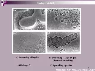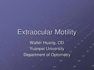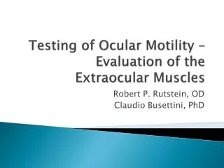
Ocular Motility
Ocular Motility. M.R Besharati MD Shahid Sadoughi University. Superior Oblique/Trochlear Muscle. Eye Muscles Left eye. Superior Rectus Muscle. Medial Rectus Muscle. Lateral Rectus Muscle. Inferior Rectus Muscle. Inferior Oblique Muscle. Anatomy Of The EOM’s. What are the actions of

Ocular Motility
E N D
Presentation Transcript
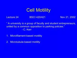
Ocular Motility M.R Besharati MD Shahid Sadoughi University
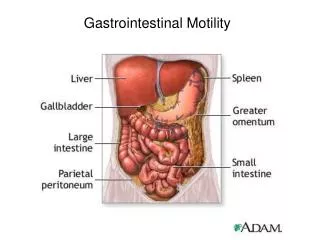
Superior Oblique/Trochlear Muscle Eye Muscles Left eye Superior Rectus Muscle Medial Rectus Muscle Lateral Rectus Muscle Inferior Rectus Muscle Inferior Oblique Muscle
Anatomy Of The EOM’s What are the actions of EOM surround each eye:
Medial Rectus • Adduction
Lateral Rectus • Abduction
Superior Rectus Elevation, Adduction, Intorsion
Inferior Rectus Depression, Adduction, Extorsion
Superior Oblique Intorsion, Depression, Abduction
Inferior Oblique Extorsion Elevation Abduction
Anatomy Of The EOM’s The two Oblique are Abductors The two Recti are Adductors The two Superiors are Intorters The two Inferiors are Extorters
Anatomy Of The EOM’s Origin A common tendinous ring (annulus of Zinn)
Anatomy Of The EOM’s Blood supply Each muscle is supplied by two Anterior Ciliary Arteries except the Lateral Rectus which is only supplied by one.
Anatomy Of The EOM’s Nerve supply Third: LPS, MR, IR, SR, IO Fourth: SO Sixth: LR
Ocular motility CN III CN IV CN III CN VI CN III CN III
Eye movement Three directions of eye movement Vertically Upward SR & IO Downward IR & SO Horizontally Abduction LR Adduction MR Torsionally Intorsion (rotate nasally) SO Extorsion (rotate temporally) IO
Ocular motility Agonist Muscles: Receive equal innervation to ensure coordinated eye movements Agonist/Antagonist Pairs (within each eye) Receive reciprocal innervation
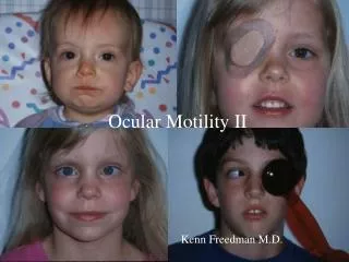
Amblyopia: History • “When the doctor sees nothing and the patient sees nothing, the diagnosis is amblyopia.”
What’s Amblyopia? • Sometimes called “lazy eye”: characterized by: • Reduced visual acuity in an otherwise normal eye. • Onset early in life (typically before age 6) • Associated with a history of abnormal binocular visual experience.
Amblyopia • Unilateral or less commonly, bilateral reduction of best corrected visual acuity that can not be attributed directly to the effect of any structural abnormality of the eye or the posterior visual pathway. Defect of central vision
Amblyopiascreening Prevalence: 2%-4% . Commonly unilateral Nearly all amblyopic visual loss is preventable or reversible with timely detection and appropriate intervention. Children with amblyopia or at risk for amblyopia should be identified at a young age when the prognosis for successful treatment is best. Role of screening is important
Amblyopia: Definition • Uncorrectable, decreased vision in an otherwise structurally normal eye • definition includes an operated eye made “structurally normal” by surgery (e.g. post cataract surgery) • May be unilateral (most common) or bilateral
Associated (causative) Conditions: • Amblyopia is generally accompanied by: • strabismus, • Anisometropia • Isoametropia • form deprivation • Occlusive
Strabismus refers to an eye-turn. • normal esotropia F F F F
Anisometropic Amblyopia e.g., one eye in focus (emmetropic) and the other out of focus (e.g. hyperopic) Amblyopia usually seen with hyperopic anisometropia
Amblyopia Functional reduction in visual acuity of an eye caused by disuse/misuse during the critical period of visual development • Strabismic Amblyopia – results from abnormal binocular interaction • The visual cortex suppresses the image from one eye • Long term suppression results in loss of vision
Amblyopia Amblyopia is the unilateral or bilateral decrease of Vision caused by form vision deprivation and/or abnormal binocular interaction for which there is no obvious cause found by physical examination of the eye. Can become irreversible if not treated before age 6 to 10 years
Management First address vision impairment caused by amblyopia Prescription of glasses to correct refractive errors Occlusion therapy Alignment Medical Glasses with/without prisms Patching Visual training exercises Surgical
Occlusion Therapy Patching the eye with the better vision Full or part-time Dependant on age/cause/severity Forces use of amblyopic eye Improvement of V.A
Why We Treat 1- Restore Stereopsis 2- Prevent Amblyopia 3- Prevent Confusion and Diplopia 4- Appearance
Hirschberg Test • Used as an initial screen for strabismus • How it works: • Stand several feet in front of child with penlight shining at eyes • Light reflection will be at the same point in each eye Normal Exotropia Esotropia
Cover Test Child fixes on target (near or far) Examiner covers one eye while observing the opposite eye for movement No movement = normal ocular alignment Uncovered eye shifts to re-fixate on object = Manifest strabismus Indicates that the covered eye was the fixating eye
Cover-Uncover Test • Used to detect latent strabismus • Child fixes on object (near or far) • A cover is placed over one eye for a few seconds then rapidly removed • The eye under the cover is observed for movement
Cover – Uncover test Orthophoria, normal No complaints, asymptomatic
Cover – Uncover test Esophoria, abnormal, common Only seen when eye is covered Often asymptomatic, no complaints
Cover – Uncover test Exophoria, abnormal, common Only seen when eye is covered Often asymptomatic, no complaints.
Alternate cover test Remember to allow the pt time to fixate on the target, give them a minute. Then quickly cover the other eye to prevent the pt from regaining fusion. But do not go back and forth quickly because the pt will not have time to refixate.
Alternate Cover test Exotropia, intermittent May be visible with or without alternate cover May have intermittent diplopia, especially when tired or sick
Alternate Cover test Exotropia, Constant May be visible with or without alternate cover May or may not have constant diplopia
Cover Uncover test Left Exotropia, Constant May be visible with or without alternate cover Right eye preference
Cover Uncover test Left Exotropia, Constant May be visible with or without alternate cover Right eye preference
Normal Convergence Convergence Insufficiency
20 How much to operate… Alternate Cover test with Prism Exotropia, Constant Use prism to quantitate the deviation. Change prism power until movement is neutralized. Use this number to plan surgery
Why We Treat The main types of Amblyopia are: 1. Strabismic amblyopia results from abnormal binocular interaction where there is continued monocular suppression of the deviating eye. It is Characterized by an impairment of vision which is present even when the eye is forced to fixate.
Why We Treat 2. Anisometropic amblyopia is caused by a difference in refractive error. It results from abnormal binocular interaction from the superimposition of a focused and unfocused image or from the superimposition of large and small images from aniseikonia. 3. Deprivation Amblyopia is caused from form vision deprivation of one eye.
Why We Treat - Confusion and Diplopia DEFINITIONS 1. Visual axis is a line that passes through the point of fixation and the fovea. The normal visual axes intersect at the point of fixation. 2. Strabismus is a misalignment of the visual axes which, initially, results in confusion and diplopia. 4. Diplopia is the simultaneous appreciation of two images of one object. it results from a failure to maintain binocular vision.