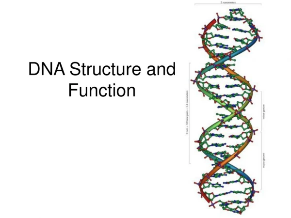
Structure and function of DNA
Structure and function of DNA. Dr. Ghada Abou El-Ella Lecturer of biochemistry Faculty of Vet. Medicine South Valley University. Central Dogma. DNA ---------→ RNA---------→Protein. This unidirectional flow equation represents the Central Dogma (fundamental law) of molecular biology.

Structure and function of DNA
E N D
Presentation Transcript
Structure and function of DNA Dr. Ghada Abou El-Ella Lecturer of biochemistry Faculty of Vet. Medicine South Valley University
Central Dogma DNA ---------→ RNA---------→Protein. • This unidirectional flow equation represents the Central Dogma (fundamental law) of molecular biology. • This is the mechanism whereby inherited information is used to create actual objects, namely enzymes and structural proteins. • An exception to the central dogma is that certain viruses (retroviruses) make DNA from RNA using the enzyme reverse transcriptase.
Gene Expression • Genes are DNA sequences that encode proteins (the gene product) • Gene expression refers to the process whereby the information contained in genes begins to have effects in the cell. • DNA encodes and transmits the genetic information passed down from parents to offspring.
Genetic code • The alphabet of the genetic code contains only four letters (A,T,G,C). • A number of experiments confirmed that the genetic code is written in 3-letter words, each of which codes for particular amino acid. • A nucleic acid word (3 nucleotide letters) is referred to as a codon.
Nucleic acids • Principle information molecule in the cell. • All the genetic codes are carried out on the nucleic acids. • Nucleic acid is a linear polymer of nucleotides
Nucleotides • Nucleotides are the unit structure of nucleic acids. • Nucleotides composed of 3 components: • Nitrogenous base (A, C, G, T or U) • Pentose sugar • Phosphate
Nitrogenous bases • There are 2 types: • Purines: • Two ring structure • Adenine (A) and Guanine (G) • Pyrimidines: • Single ring structure • Cytosine (C) and Thymine (T) or Uracil (U).
Types of Nucleic acids There are 2 types of nucleic acids: • Deoxy-ribonucleic acid (DNA) • Pentose Sugar is deoxyribose (no OH at 2’ position) • Bases are Purines (A, G) and Pyrimidine (C, T).
Ribonucleic acid (RNA) • Pentose Sugar is Ribose. • Bases are Purines (A, G) and Pyrimidines (C, U).
Linear Polymerization of Nucleotides • Nucleic acids are formed of nucleotide polymers. • Nucleotides polymerize together by phospho-diester bonds via condensation reaction. • The phospho-diester bond is formed between: • Hydroxyl (OH) group of the sugar of one nucleotide. • Phosphate group of other nucleotide
Polymerization of Nucleotides • The formed polynucleotide chain is formed of: • Negative (-ve) charged Sugar-Phosphate backbone. • Free 5’ phosphate on one end (5’ end) • Free 3’ hydroxyl on other end (3’ end) • Nitrogenous bases are not in the backbone • Attached to the backbone • Free to pair with nitrogenous bases of other polynucleotide chain
Polymerization of Nucleotides • Nucleic acids are polymers of nucleotides. • The nucleotides formed of purine or pyrimedine bases linked to phosphorylated sugars (nucleotide back bone). • The bases are linked to the pentose sugar to form Nucleoside. • The nucleotides contain one phosphate group linked to the 5’ carbon of the nucleoside. Nucleotide = Nucleoside + Phosphate group
N.B. • The polymerization of nucleotides to form nucleic acids occur by condensation reaction by making phospho-diester bond between 5’ phosphate group of one nucleotide and 3’ hydroxyl group of another nucleotide. • Polynucleotide chains are always synthesized in the 5’ to 3’ direction, with a free nucleotide being added to the 3’ OH group of a growing chain.
Complementary base pairing • It is the most important structural feature of nucleic acids • It connects bases of one polynucleotide chain (nucleotide polymer) with complementary bases of other chain • Complementary bases are bonded together via: • Double hydrogen bond between A and T (DNA), A and U (RNA) (A═T or A═U) • Triple H-bond between G and C in both DNA or RNA (G≡C)
Significance of complementary base pairing • The importance of such complementary base pairing is that each strand of DNA can act as template to direct the synthesis of other strand similar to its complementary one. • Thus nucleic acids are uniquely capable of directing their own self replication. • The information carried by DNA and RNA direct the synthesis of specific proteins which control most cellular activities.
DNA structure • DNA is a double stranded molecule consists of 2 polynucleotide chains running in opposite directions. • Both strands are complementary to each other. • The bases are on the inside of the molecules and the 2 chains are joined together by double H-bond between A and T and triple H-bond between C and G. • The base pairing is very specific which make the 2 strands complementary to each other. • So each strand contain all the required information for synthesis (replication) of a new copy to its complementary.
Forms of DNA 1- B-form helix: • It is the most common form of DNA in cells. • Right-handed helix • Turn every 3.4 nm. • Each turn contain 10 base pairs (the distance between each 2 successive bases is 0.34 nm) • Contain 2 grooves; • Major groove (wide): provide easy access to bases • Minor groove (narrow): provide poor access.
2- A-form DNA: • Less common form of DNA , more common in RNA • Right handed helix • Each turn contain 11 b.p/turn • Contain 2 different grooves: • Major groove: very deep and narrow • Minor groove: very shallow and wide (binding site for RNA) 3-Z-form DNA: • Radical change of B-form • Left handed helix, very extended • It is GC rich DNA regions. • The sugar base backbone form Zig-Zag shape • The B to Z transition of DNA molecule may play a role in gene regulation.
Denaturing and Annealing of DNA • The DNA double strands can denatured if heated (95ºC) or treated with chemicals. • AT regions denature first (2 H bonds) • GC regions denature last (3 H bonds) • DNA denaturation is a reversible process, as denatured strands can re-annealed again if cooled. • This process can be monitored using the hyperchromicity (melting profile).
Hyperchromicity (melting profile) • It is used to monitor the DNA denaturation and annealing. • It is based on the fact that single stranded (SS) DNA gives higher absorbtion reading than double stranded (DS) at wavelength 260º. • Using melting profile we can differentiate between single stranded and double stranded DNA.
SS SS Ab260 DS Tm Temperature Hyperchromicity (melting profile) Using melting profile we can differentiate between SS DNA and DS DNA • Tm (melting temp.): temp. at which 50% of DS DNA denatured to SS • Heating of SS DNA: little rise of Ab reading • Heating of DS DNA: high rise of Ab reading
SS AT rich DNA GC/AT DNA GC rich DNA DS Tm2 Tm1 Tm3 Melting profile continue….. • Melting profile can be also used to give an idea about the type of base pair rich areas using the fact that: • A═T rich regions: denatured first (low melting point) • G≡C rich regions: denatured last (higher melting point) Tm1: Small melting temp. of AT rich DNA Tm2: higher melting temp. of AT/GC equal DNA Tm3: Highest melting temp. of GC rich DNA
RNA structure • It is formed of linear polynucleotide • It is generally single stranded • The pentose sugar is Ribose • Uracile (U) replace Thymine (T) in the pyrimidine bases. Although RNA is generally single stranded, intra-molecular H-bond base pairing occur between complementary bases on the same molecule (secondary structure)
Types of RNA • Messenger RNA (mRNA): • Carries genetic information copied from DNA in the form of a series of 3-base code, each of which specifies a particular amino acid. • Transfer RNA (tRNA): • It is the key that read the code on the mRNA. • Each amino acid has its own tRNA, which binds to it and carries it to the growing end of a polypeptide chain. • Ribosomal RNA (rRNA): • Associated with a set of proteins to form the ribosomes. • These complex structures, which physically move along the mRNA molecule, catalyze the assembly of amino acids into protein chain. • They also bind tRNAs that have the specific amino acids according to the code.
RNA structure • RNA is a single stranded polynucleotide molecule. • It can take 3 levels of structure; • Primary: sequence of nucleotides • Secondary: hairpin loops (base pairing) • Tertiary: motifs and 3D foldings
RNA structure Transfer RNA (tRNA) structure
DNA Replication • Replication of the DNA molecule is semi-conservative, which means that each parent strand serves as a template for a new strand and that the two (2) new DNA molecules each have one old and one new strand. • DNA replication requires: • A strand of DNA to serve as a template • Substrates - deoxyribonucleoside triphosphates (dATP, dGTP, dCTP, dTTP). • DNA polymerase - an enzyme that brings the substrates to the DNA strand template • A source of chemical energy to drive this synthesis reaction.
DNA Replication • Nucleotides are always added to the growing strand at the 3' end (end with free -OH group). • The hydroxyl group reacts with the phosphate group on the 5' C of the deoxyribose so the chain grows • Energy is released when the bound linking 2 of the 3 phosphate groups to the deoxyribonucleoside triphosphate breaks • Remaining phosphate group becomes part of the sugar-phosphate backbone
Step 1 - Unwinding and Exposing Strands • DNA strands are unwound and opened by enzymes called HELICASES • Helicases act at specific places called ORIGINS OF REPLICATION • Synthesis of new DNA strands proceeds in both directions from an origin of replication resulting in a bubble with REPLICATION FORKS at each growing point.
Step 2 - Priming the Strand • In order to begin making a new strand, a helper strand called a PRIMER is needed to start the strand. • DNA polymerase, an enzyme, can then add nucleotides to the 3' end of the primer. • Primer is a short, single strand of RNA (ribonucleic acid) and is complimentary to the DNA template strand. • Primers are formed by enzymes called PRIMASES.
Step 3 - Strand Elongation • DNA Polymerase III catalyses elongation of new DNA strands in prokaryotes • Two molecules of DNA polymerase III clamp together at the replication forks, each acting on 1 of the strands • One strand exposed at its 3' end produces a daughter strand which elongates from its 5' to 3' end and is called the LEADING STRAND. This strand is synthesized continuously and grows from 5' to 3'.
Step 3 - Strand Elongation • The second daughter strand is called the LAGGING STRAND and is antiparallel to the leading strand. It’s template is exposed from the 5' to 3' end but it must direct the 5' to 3' synthesis of the lagging strands, since nucleotides are added at the 3' end of the chain. • The lagging strand is constructed in small, backward directed bits consisting of discontinuous sections of 100-200 nucleotides in eukaryotes and 1000-2000 nucleotides in prokaryotes, called OKAZAKI FRAGMENTS.
Step 3 - Strand Elongation • When an Okazaki fragment forms: DNA polymerase I removes the RNA primer and replaces it with DNA adjacent to the fragment. • leaving 1 bond between adjacent fragments missing. • A second enzyme called a DNA LIGASEcatalyses the formation of the final bond.
Telomerase • Telomerase is a reverse transcriptase that contain an RNA template, adds nucleotides to the 3’end of the lagging-strand template and thus prevents shortening of lagging strands during replication of linear DNA molecules such as those of eukaryotic chromosomes.