Download
1 / 26
280 likes | 676 Vues
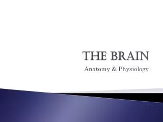
Brain anatomy & physiology and Neurological Assessment. James Bitmead (Clinical Practice Facilitator, UCLH) Angela Roots (Practice Development Nurse, GSTT). What is a stroke?.

E N D
Brain anatomy & physiologyand Neurological Assessment James Bitmead (Clinical Practice Facilitator, UCLH) Angela Roots (Practice Development Nurse, GSTT)
What is a stroke? • interruption of the blood supply to the brain, caused by a blocked or burst blood vessel…cuts off the supply of oxygen and nutrients, causing damage to the brain tissue. (World Health Organisation 2010)
Aetiology of Stroke • Cerebral infarction/ischaemic 81% • Intracerebral haemorrhage 13% • Subarachnoid haemorrhage 6% • Risk of recurrence within 5 years 30-40% (Stroke Association 2010)
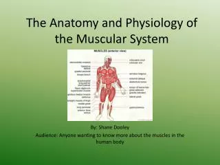
Speech centres • Broca; control the muscles of the larynx, pharynx and mouth that enable us to speak • Wernicke’s area, injury here may result in receptive dysphasia.
Ischemic stroke (Thrombo/embolic stroke) • hypercholesterolemia • hypertension • Atrial fibrillation • Ischaemic heart disease/angina • Peripheral vascular disease • Diabetes
Previous stroke/TIA • Smoking • Increased alcohol intake • Poor diet/obesity • Increased age-atherosclerosis • Oral Contraceptive Pill • Drug misuse
Haemorrhagic Stroke • Chronic high blood pressure. • Amphetamine. • Amyloid angiopathy • Arterial Venous malformation (AVM), • inflammation of blood vessels (vasculitis), • bleeding disorders, • anticoagulants,
Early Signs Agitation Vomiting Headache Dilated pupils Later Signs Increased systolic blood pressure Bradicardia Abnormal respiratory pattern Raised Intracranial Pressure
Causes Oedema Haemorrhage Tumour Encephalopathy Treatment Steroids Manitol Hyperventilation Hemicraniectomy Causes and Treatment
Neurological Assessment • AVPU – what does this mean? • Blood sugar • Pupils • Then move onto GCS and full neuro assessment
The Glasgow Coma Scale • The eye opening category is performed once the patient is fully awake not before • The verbal category means a verbal response – the patient has to verbally indicate their orientation to time, place and person to be orientated • Mute dysphasic patients cannot score 5 on the verbal category
The Glasgow Coma Scale • The motor response is best done without the patient copying your action – truly obeying command not copying! • Score the GCS in your documentation as GCS=15 E 4 V 5 M6
MRC limb power grading • 5= full strength • 4=able to move against resistance but easily overcome • 3= able to move against gravity but not resistance • 2= able to move but not against gravity • 1= flicker • 0= no movement
Neurological assessment • Score the patient as you see them – no guessing or backdating the results • If they do not meet one criteria move down the score to the next one • Always start the assessment with the patient as awake as possible (even at 2am)
Changing GCS • If patient looks different to the GCS scoring do a set of obs together at hand over • Consistency with using the neuro. Obs is vital to detecting changes in the patients • Don’t forget to spot other changes like increasing confusion even if the GCS hasn’t yet changed
Patterns of change in GCS • Dropping obviously! • Fluctuating widely – could it represent seizure (sub-clinically) • Increasing difficulty in obtaining the same GCS • Small changes within the category – e.g. confused but worsening confusion, obeys some commands but not others • Vital signs changes- will come to later
ESCALATE!!!! If you are concerned at all, do not be afraid to escalate!!!!! • Band 6 • Site Nurse Practitioner • Consultant oncall