CASE OF THE MONTH
250 likes | 553 Vues
CASE OF THE MONTH. Submitted by: Dr. Cecil White Jr. HISTORY. 32 year-old female; Medical history within normal limits. OMFS performed LeFort I osteotomy in maxilla (w/BSSO in mandible). The mesial root of #6 was cut, and developed a persistent radiolucent area; no pain involved.

CASE OF THE MONTH
E N D
Presentation Transcript
CASE OF THE MONTH Submitted by: Dr. Cecil White Jr.
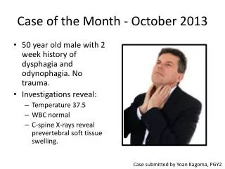
HISTORY • 32 year-old female; Medical history within normal limits. • OMFS performed LeFort I osteotomy in maxilla (w/BSSO in mandible). • The mesial root of #6 was cut, and developed a persistent radiolucent area; no pain involved. • #6 developed draining fistula w/no response to electric pulp tester. Subsequent pain upon mastication, 6 months later, prompted non-surgical endo to #6. • Referral to periodontist, 54 months following original orthognathic surgery.
CLINICAL EXAM • 10 mm probing depth (PD) at mesial of #6, and 7 mm PD at distal of #7. • Miller’s Class I mobility of #6 and #7. • #6 displayed purulence and bleeding upon probing. • Radiographs displayed radiolucent area at mesial of #6, with partial loss of root structure in apical 1/3rd of tooth.
TREATMENT PLAN • Phase I: • Scaling/root planing (SRP) of area performed. • Reevaluation at 8 weeks following SRP revealed persistent problem, with no improvement in probing depth/clinical attachment levels. • Phase II: • Open flap debridement to assess lesion/defect
INTRA-OPERATIVE FINDINGS • Combination 1- and 2-wall intrabony defect, extending from the mesial surface, to the distopalatal line angle of #6. • Vertical dimension of the defect ranged from 4-6 mm. • A 5 mm x 2 mm segment of gutta-percha was exposed, starting 4 mm apical to the CEJ.
POTENTIAL TREATMENT OPTIONS • Application of Enamel Matrix Derivative (EMD) to defect and root surface of #6, closure. • Extract tooth #6, immediate implant to #6, bone graft/barrier, closure. • Glass ionomer cement to root surface defect, root surface conditioning with tetracycline (TCN), bone graft/TCN/barrier, closure. • Extract teeth #6 and #7, bone graft/barrier, closure; subsequent implant placement. • Extract #6, bone graft/barrier; subsequent placement of Fixed Bridge (#4 - #8).
CHOSEN TREATMENT OPTION Glass ionomer cement to root surface defect, root surface conditioning with tetracycline (TCN), bone graft/TCN/barrier, closure.
DFDBA graft/tetracycline combination placed into defect, following root surface conditioning with tetracycline and glass ionomer cement repair of root surface defect.
Barrier removal at 6 weeks, with “regenerative” soft tissue present
Re-entry of #6 area at 24 months following original open flap/root repair/bone graft procedure (i.e. “original” surgery)
Restoration at 30 months following “original” surgery/6 months following “re-entry” procedure - Facial
Restoration at 30 months following “original” surgery/6 months following “re-entry” procedure - Palatal
PRE-SURGERY POST-SURGERY/RESTORATIVE
PRE-SURGERY 24 MONTH POST-SURGERY
SUMMARY • PROBLEM • Chronic periodontal lesion, with root surface defect, caused by errant section of root surface during orthognathic surgery procedure. • TREATMENT • Situation was treated with open flap debridement, repair of root surface defect with glass ionomer cement, root surface conditioning with tetracycline, combination DFDBA/tetracycline (4:1 ratio), and placement of e-PTFE barrier. • OUTCOME • 1 to 3 mm probing depths, no mobility, and no bleeding on probing to sites #6 and #7.