
Understanding the Gastrointestinal System and Its Role in Digestion
The gastrointestinal system is essential for processing food and extracting nutrients. It includes various functions such as ingestion, mastication, digestion, secretion, absorption, and elimination of waste. The mouth, containing salivary glands and teeth, initiates digestion through mechanical and chemical processes. Saliva plays a crucial role in breaking down carbohydrates and maintaining oral hygiene. Additionally, dental health is vital as issues like cavities and periodontal disease can significantly affect overall well-being. This chapter delves into the anatomy, functions, and common disorders associated with the gastrointestinal system.

Understanding the Gastrointestinal System and Its Role in Digestion
E N D
Presentation Transcript
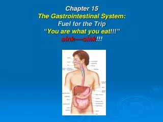
Chapter 15The Gastrointestinal System:Fuel for the Trip“You are what you eat!!!”oink----oink!!!
Introduction Gastrointestinal System Functions: • Ingest raw materials • Physically & chemically digest raw material to usable elements. • Absorb elements • Eliminate what is NOT useable
System Functions • Ingestion: food enters mouth • Mastication: chewing • Digestion: chemical act of breaking down food into small molecules. • Secretion: acids, buffers, enzymes, & H2O aid in breakdown of food. • Absorption: molecules pass through lining of digestive tract. • Excretion or defecation: elimination of waste products.
Buccal (oral) Cavity • Lips: act as door to cavity • Hard & Soft Palate: form roof of mouth • Tongue: acts as floor • Cheeks: form walls
Tongue • Muscle: provides taste stimuli to brain, determines temperature, manipulates food, & aids in swallowing. • Saliva: added to moisten & soften food, while teeth crush food. • Bolus: ball-like mass, pushed by tongue so it may be swallowed, passed to pharynx. • Lingual Frenulum: membrane under tongue, keeps you from swallowing tongue & aids in speaking.
Salivary Glands • Sublingual: found under tongue • Submandibular: located along both sides of inner surface of mandible, or lower jaw. • Controlled by: autonomic nervous system • Parotid: slightly inferior & anterior to each ear. Swell with “viral Parotitis..”
Salivary Glands con’t • Produces: 1–1.5 liters of saliva QD • Keep mouth moist: but idea or presence of food increase production significantly. • Contains: 99.4% water, & contains antibodies, buffers, ions, waste products, & enzymes.
Salivary Glands con’t • Enzymes: act as organic catalysts to speed up chemical reactions. • Salivary Amylase: speeds chemical activity of breaking down carbohydrates. • Saliva cleans oral surfaces, reducing amount of bacteria that grows in mouth.
Teeth • Deciduous: first set of teeth as a baby • First tooth: appears @ 6 months of age; lower central incisors appear first, all 20 teeth in place by age 2½. • Between 6 and 12 years these teeth fall out, are replaced by 32 permanent teeth. • Wisdom teeth: appear by age 21
Teeth Con’t • Incisors: at front of mouth, blade shaped, used to cut food. • Canine: for holding, tearing, or slashing food; known as eyeteeth or cuspids, located next to incisors. • Bicuspids: or premolars: transitional teeth • Molars: have flattened tops; both bicuspids & molars are responsible for crushing & grinding food.
Parts of Tooth: Crown: covered by hard enamel. Neck: transitional section that leads to root. Root: nestled in bony socket, held in place by fibers of periodontal ligament. Dentin: made of mineralized bone-like substance. Teeth Con’t
Connective tissue: pulp, located in pulp cavity Pulp cavity: contains blood vessels & nerves providing nutrients & sensation; nerves & blood vessels get to pulp cavity via root canal. Cementum: (soft version of bone) covers dentin of root, aiding in securing periodontal ligament. Teeth Con’t
TeethCon’t • Gingiva: gums, help hold teeth in place • Epitheal cells form tight seal around tooth to prevent bacteria from coming into contact with tooth’s cementum.
Pathology Connection: Oral Disorders Dental Caries (cavities) • Form whenmicroorganisms attack tooth enamel • Related to dental plaque: sticks to teeth forming sticky substance. • Forms great hideout for bacteria • Bacteria createsacids that attack surface of teeth.
Risk Factors for Plaque Formation • High carbohydrate diet • Poor dental hygiene • Lack of regular visits to dentist
Risk Factors for Plaque Formation con’t RX: • Clear out & fill caries • Rx infection Prevention: • Proper dental care • Fluoride in H2O & tooth paste • Evaluate for heart disease & buccal ca
Pathology Connection:Periodontal Disease • Plaque & bacteria affects gums & supportive structures of teeth. • Can result ingingivitis, bleeding & tooth loss
Pathology Connection:Oral & Lip Cancer Cause: • Excessive sun exposure • Tobacco • ETOH
Oral & Lip Cancer con’t Lukoplakia: • white patch of tissue in mouth • associated with use of chewing tobacco
Pathology Connection:Stomatitis • Inflammation of oral mucosa • poor fitting dentures • Apthousstomatitis (“canker sores”) • Cheilitis: cracking & inflammation of lips & corners of mouth; often related to infection, allergy, or nutritional deficiency.
Pharynx (3 Parts) Nasopharynx: • primarily part of respiratory system, blocked by soft palate. Oropharynx & laryngopharynx: • act as passageway for food, water, & air; epiglottis covers trachea to prevent food from entering lungs, forcing food into opening for esophagus.
Esophagus • 10 inches long, is connected to stomach • from pharynx, through thoracic cavity, through diaphragm, connecting to stomach in peritoneal cavity. • normally collapsed tube until “bolus” of food swallowed. • Peristalsis: pushes food down esophagus
Esophagus con’t • lined withstratified squamous epithelium that secrete mucus to make walls slippery; cells make lining. • resistant toabrasion, temperature extremes, & irritation. • Pharyngoesophageal sphincter: relaxes to open esophagus so food can enter.
Esophagus con’t • Lower Esophageal Sphincter: opening door to stomach & closing to prevent acidic gastric juices from splashing into esophagus causing heartburn. • process of swallowing: food9 seconds; fluid take only seconds to reach stomach.
Located: ULQ under diaphragm, posterior to Liver. 10 inches long with diameter dependent on how much just eaten. 4 liters when filled Rugae: folds, help stomach expand and contract. Stomach
4 Function of Stomach • Holdingarea for received food • Chemical digestion: gastric acids & enzymes mix with food. • Regulates rate of Chyme movement into small intestines. • Absorbs small amounts of H2O & ETOH
How Fast Stomach Empties • 4 hours to empty following meal • Liquids & carbohydrates pass quickly • Proteins take longer • Fats take longest 4-6 hours
Cardiac Region: surrounding lower esophageal sphincter. Fundus: laterally & slightly superior to cardiac region. Temporarily holds food as it enters stomach. Body: Mid-portion Pylorus: 1. terminal end of stomach 2. most of work performed 3. where food passes through pyloric sphincter into small intestine. 4 Regions of Stomach
Chemical Digestion Gastric Juice: • 1500 mls produced QD • hydrochloric acid (HCl) • pepsinogen • mucus
Pepsinogen, HCL & Pepsin Enzymes • chief digestive enzyme • secreted bychief cells • HCL secreted by parietal cells combining to produce pepsin. • Pepsin breaks down protein • HCl breaks down connective tissue
Stomach & Enzymes con’t • HCL: pH of 1.5–2.0, effective at killing pathogens. • Mucous cells: generate thick layer of mucus shielding stomach from effects of stomach acids. • Stomach secretes intrinsic factor, allowing vitamin B12 to be absorbed. • Enzyme activity controlled by parasympathetic nervous system (vagus nerve) • Vagus increases motility & secretory rates of gastric glands.
I. Cephalic Phase: sensory stimulation (sight or smell of food) stimulates parasympathetic nerves via medulla oblongata Gastrin released stimulating gastric gland activity in stomach 3 Phases of Gastric Juice Production
3 Phases of Gastric Juice Production con’t II. Gastric Phase: • 2/3 of gastric juices secreted as food enters stomach & distends walls. • signaling stomach to secrete more gastric fluid
3 Phases of Gastric Juice Production con’t III. Intestinal Phase: • food enters duodenum, distending & sensing acidity. • intestinal hormones released • slowing gastric gland secretions • lasts until bolus leaves duodenum
Rate of Movement of Chyme If too slow: • rate of nutrient digestion & absorption decreased • may allow acidity of chyme to cause erosions of stomach lining (ulcers). If too quick: • food particles may not be sufficiently mixed with gastric juices. • insufficient digestion; chyme not given time to neutralize can cause erosion of intestinal lining (ulcers).
Pathology Connection: Stomach Acid Disorders • Gastroesophageal Reflux Disease (GERD) • Condition where acidic stomach contents “squirt” back into esophagus • Since esophagus does not have protective mucus, can cause inflammation and ulceration of esophageal tissue • Scar tissue can eventually form, causing narrowing of esophagus • If left untreated, constant inflammation can lead to esophageal cancer
GERD cont. • s/sepigastric pain and burning, can be worse when lying down • d/x symptoms, upper GI • R/x • Antacids: treat burning sensation by decreasing acid • Acid reducing meds • Lifestyle changes: may help prevent GERD • Limiting fats, alcohol, caffeine and chocolate in diet • Avoiding smoking • Avoiding lying down in 4 hours after eating • Sleeping with head of bed elevated • If obese, weight loss
Peptic Ulcers Etiology: Breakdown of mucosal membrane in esophagus, stomach, or small intestine; develop most commonly in duodenum Factors that increase risk: • Helicobacter pylori (H. pylori) infection in stomach: • Smoking • Heavy/chronic alcohol consumption • Use of NSAID medications (including aspirin and others) • Caffeine consumption
Peptic Ulcer cont. • Use of corticosteroid medications • Stress
Small Intestine • major organ of digestion, is where most of food digested • average length of 6–20 feet and diameter ranging from 2.5-4 cm • Walls secrete digestive enzymes and hormones to stimulate pancreas
Small Intestine cont. • 80% of absorption of usable nutrients occurs in sm. Intestine • Remaining 20% absorbed in stomach • Any residue not utilized in small intestine sent to large intestine for removal from body
Sections of sm. intestine • Three regions • Duodenum: approximately 25 cm long (10 inches) • Jejunum: middle section, approximately 2.5 m long • Ileum: terminal end, 2 meters long, attaches to large intestine at ileocecal valve
Sm. Intestine cont. • Pyloric valve allows small portions of chyme to enter duodenum • Pancreas and gallbladder add secretions: bile from gallbladder, pancreatic juice with enzymes from pancreas • Bile emulsifies fat, making fat disperse in water • Pancreatic juice contains sodium bicarbonate which neutralizes acidic chyme


