BRAIN DAYS PART III
BRAIN DAYS PART III . DIENCEPHALON. IN 10 QUESTIONS. Kaan Yücel M.D.,Ph.D. Learning Objectives Explain the components of the diencephalon Explain the anatomical features of the structures in diencephalon. 1. Diencephalon. Prosencephalon [ Forebrain ] Telencephalon Diencephalon.

BRAIN DAYS PART III
E N D
Presentation Transcript
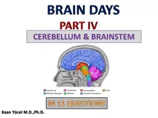
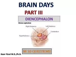
BRAIN DAYS PART III DIENCEPHALON IN 10 QUESTIONS Kaan Yücel M.D.,Ph.D.
Learning Objectives Explainthecomponents of thediencephalon Explaintheanatomicalfeatures of thestructures in diencephalon
1. Diencephalon Prosencephalon [Forebrain] Telencephalon Diencephalon
2. Diencephalon is composed of… (1) thalamus (2) subthalamus [-sub: 'inferior to”] (3) epithalamus [-epi: “superior to”] (4) hypothalamus [-hypo: “under” ]
2. Diencephalon extends... posteriorly where 3rd ventricle becomes continuous with the cerebral aqueduct anteriorly interventricular foramina Amidline structure with symmetrical right and left halves
2. Diencephalon’s inferior surface • Only area exposed to the surface in the intact brain. • Formed by hypothalamic and other structures, which include, from anterior to posterior: • optic chiasma • infundibulum • with the tuber cinereum[x] • mammillary bodies [x]
2. Diencephalon’s superior surface • Concealed by the fornix. • The actual superior wall of the diencephalon is formed by roof of the third ventricle. • A pair of vascular processes, choroid plexuses of the third ventricle, project downward from the midline into the cavity of the third ventricle and CSF is produced here.
2. Diencephalon’s lateral surface • Bounded by the internal capsule • consists of nerve fibers • connect the cerebral cortex with parts of the brainstem and spinal cord.MRI of the brain, T1-weighted axial cut 1.Insula 2. Internal capsule 3.Caudate nucleus 4.Putamen 5.Internal capsule Posterior limb 6.Splenium, corpus callosum 7.Thalamus
2. Diencephalon’s medial surface a.k.a. Lateral wall of the third ventricle Superior part by medial surface of thalamus hypothalamic sulcus Inferior party byhypothalamus • striamedullaris thalami • Afferent fibers to the habenular nucleus • Forms a ridge along the superior margin of the medial surface of the diencephalon
3. Thalamus L. thalamus "inner chamber," from Gk. thalamos "inner chamber, bedroom" Large ovoid mass of gray matter that forms the major part of the diencephalon. On each side of the third ventricle.
3. Thalamus The superior surface of the thalamus is covered medially by the telachoroidea and the fornix The inferior surface is continuous with the tegmentum of the midbrain.
3. Thalamus The medial surface of the thalamus forms the superior part of the lateral wall of the third ventricle Usually connected to the opposite thalamus by a band of gray matter, interthalamic connection (interthalamic adhesion-adhesiointerthalamica-massainterrmedia) Found in 70-80% of humans Axial T2-weighted MR image C = caudate nucleus G = globuspallidus L = lentiform nucleus (G+P) P = putamen T = thalamus
Important station that receives the main sensory tracts (except the olfactory pathway). • A station where much of the information is integrated and relayed to the cerebral cortex and many other subcortical regions. • A key role in the integration of visceral and somatic functions.
3. Thalamus Anterior thalamic nuclei, which receive the mammilothalamic tract from the mammillary nuclei. Associated with of that of the limbic system and is concerned with emotional tone and the mechanisms of recent memory.
3. Thalamus Medial part of the thalamus large dorsomedial nucleus and several smaller nuclei. 2 connections with the whole prefrontal cortex and hypothalamic nuclei. Responsible for integration of a large variety of sensory information relation of this information to one’s emotions
Striaterminalis: Runs @ lateralmargin of thalamus Fromamygalatohypothalamus Majoroutput of amygdala
4. Hypothalamus CHIEF OF THE HORMONES from the region of the optic chiasma to the caudal border of the mammillary bodies
4. Hypothalamus four distinct groups in the rostral-caudal plane of the third ventricle: preoptic(above and in front of the optic chiasm - telencephalicextension of the basal forebrain, but functionally diencephalonic) chiasmatic(above and around the optic chiasm) tuberal(above and around the "tuber cinereum", i.e. pituitary stalk) posterior region which includes the mammillary bodies.
4. Hypothalamus When observed from below, the hypothalamus is seen to be related to the following structures, from anterior to posterior: optic chiasma tuber cinereum& infundibulum mammillary bodies
5. Subthalamus Inferiortothalamus Betweenthalamus-midbrain Anteriomedially- hypothalamus Importantconnectionswiththestriatum Fxn: Control of muscleactivity
Adhesio interthalamica Septumpellicidum Pinealgland Anteriorcommissure Posteriorcommissure Corporaquadrigemina AC Thalamus Opticchiasm Hypothalamus Mamillary body Cerebralaqueduct Pituitarygland
6. Pituitarygland SIDEKICK OF THE HYPOTHALAMUS
7. TuberCinereum Interthalamicadhesion
8. Mammillarybodies Twosmall hemispherical bodies situated side by side posterior to the tuber cinereum. Possessa central core of gray matter invested by a capsule of myelinated nerve fibers. Korsakoff’sSyndrome Wernicle-KorsakoffSyndrome CopenhaverBR, Rabin LA, Saykin AJ, Roth RM, Wishart HA, Flashman LA, Santulli RB, McHugh TL, Mamourian AC.Thefornixandmammillarybodies in olderadultswithAlzheimer'sdisease, mildcognitiveimpairment, andcognitivecomplaints: a volumetric MRI study. PsychiatryRes. 2006 Oct 30;147(2-3):93-103. YoneokaY, Takeda N, Inoue A, Ibuchi Y, Kumagai T, Sugai T, Takeda K, Ueda K.AcuteKorsakoffsyndromefollowingmammillothalamictractinfarction. AJNR Am J Neuroradiol. 2004;25(6):964-968. Vann SD. Re-evaluating the role of the mammillary bodies in memory.Neuropsychologia. 2010;48(8):2316-2327.
Septumpellicidum Fornix Thalamus Hypothalamus AC PC Optic chiasma Mammillarybodies Pituitary gland
Fornix Interthalamic adhesion Laminaterminalis Thalamus Habenula AC Hypothalamus Opticchiasm Infundibulum Pinealgland Pituitarygland Tubercinerum PC
8. Epithalamus Pinealgland+habenularnucleus+striamedullaristhalami Pinealgland: Inhibition of hormones Circadianrhythm day & nigh time Melatonin Habenula: potential importance of the lateralhabenularnucleus in regulating the dopamine reward signal Haber SN, Knutson B. The reward circuit: linking primate anatomy and human imaging.Neuropsychopharmacology. 2010;35(1):4-26.
9. Third ventricle Betweentwothalami LateralVentricle Interventricularforamina (foramina of Monro) 4th Ventricle Cerebralaqueduct (of Sylvius)
9. Third ventricle Anteriorwall Laminaterminalis AC Posteriorwall Cerebralaqueduct Superiorto PC pinealrecess Superiortopinealrecesshabenularcommissure Lateralwall Sup:Medialsurface of thalamus Inf: Hypothalamus Superiorlylimitedby striamedullaristhalami.
Anterior commissure (AC) Connecting temporal lobes Posterior commissure (PC) Pupillary eye reflex