Download
1 / 29
290 likes | 563 Vues
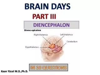
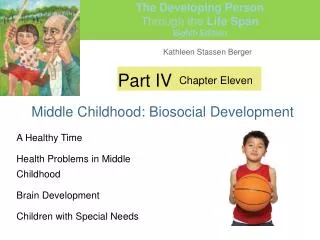
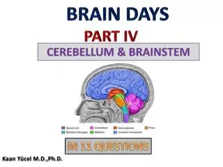
BRAIN DAYS PART IV . CEREBELLUM & BRAINSTEM. IN 11 QUESTIONS. Kaan Yücel M.D.,Ph.D. Learning Objectives Explain the anatomical structures in the brainstem Explain the parts of the cerebellum. Cerebellum L. Little brain.

E N D
BRAIN DAYS PART IV CEREBELLUM & BRAINSTEM IN 11 QUESTIONS Kaan Yücel M.D.,Ph.D.
Learning Objectives Explaintheanatomicalstructures in thebrainstem Explaintheparts of thecerebellum
Cerebellum L.Little brain • Largest part of the hindbrain [medulla, pons, and cerebellum] • Lies posterior to the fourth ventricle, the pons, and the medulla oblongata. • Situated in the posterior cranial fossa
Cerebellum L.Little brain • Covered superiorly by the tentoriumcerebelli. • Made up of two lateral cerebellar hemispheres and a median vermis (L. “worm”). • The surface displays slender and parallel elevations (ridges) known as folia and depressions (grooves) known as sulci that facilitate a great increase in the surface area of the cerebellar cortex.
Cerebellum Cerebellar hemispheres Lobe Anterior lobe primary fissure Posterior lobe uvulonodular fissure Flocculonodular lobe Lobules
Cerebellum Superiorview Inferiorview
Cerebellar Peduncles The cerebellum is linked to other parts of the central nervous system by numerous efferent and afferent fibers that are grouped together on each side into three large bundles, or peduncles. Superiorcerebellar peduncles connect the cerebellum to the midbrain Middlecerebellar peduncles connect the cerebellum to the pons Inferiorcerebellar peduncles connect the cerebellum to the medulla oblongata.
Non-motor functions of the cerebellum • Cerebellum is critical for many functions other than thecoordination of movement. • Engaged also in the regulation of cognition and emotion. • Cerebellar lesions can also result in the cerebellar cognitive affective syndrome, including executive, visual-spatial, and linguistic impairments, and affective dysregulation.
BRAINSTEM Nuclei of 12 cranialnerves 10 of them in thebrainstem midbrain pons medulla Of the IV & III Of theother 4 VIII,VII,VI,V Of thelast 4 XII,XI,X, IX
Midbrain Tectum - roof Tegmentum- cover PosteriorpartTectum superior colluculi visual reflexes inferior colluculi lower auditory centers corpora quadrigemina AnteriorpartTegmentum Belowthecerebralaqueduct
Midbrain The midbrain comprises two lateral halves cerebral peduncles anterior part: crus cerebri substantianigra posterior part: tegmentum
Substantianigra • Largemotor nucleus between tegmentum & crus cerebri • Concernedwith muscle tone • Connectedto the cerebral cortex, spinal cord, hypothalamus, and basal nuclei.
Medulla [oblongata] • In the posterior cranial fossa, lying beneath the tentoriumcerebelli and above the foramen magnum. • Related anteriorly to the basal portion of the occipital bone and the upper part of the odontoid process of the axis and posteriorly to the cerebellum.
Medulla oblongata • Not only contains many cranial nerve nuclei that are concerned with vital functions (e.g., regulation of heart rate and respiration), but it also serves as a conduit for the passage of ascending and descending tracts connecting the spinal cord to the higher centers of the nervous system
Reticularformation • The reticular formation (L. reticulum, “little net”) consists of various distinct populations of cells embed in a network of cell processes occupying the central core of the brainstem. • The reticular formation and the olfactory and limbic systems are interrelated as a result of their participation in visceral functions and behavioral responses.
Reticularformation More than 100 nuclei scattered throughout the tegmentum of the midbrain, pons and medulla have been identified as being part of the brainstem reticular formation.
Reticularformation 1- The regulation of the level of consciousness, and ultimately cortical alertness 2- The control of somatic motor movements 3- The regulation of visceral motor or autonomic functions 4- The control of sensory information
Autonomic Nervous System SympatheticParasympathetic Anatomicaldifferences, differences in the neurotransmitters, differences in the physiologic effects The autonomic nervous system and the endocrine system control the internal environment of the body. The various activities of the autonomic and endocrine systems are integrated within the hypothalamus.
Autonomic Nervous System Sympatheticpart prepares and mobilizes the body in an emergency, when there is sudden severe exercise, fear, or rage. Parasympatheticpart aims at conserving and storing energy, in the promotion of digestion and the absorption of food by increasing the secretions of the glands of the gastrointestinal tract and stimulating peristalsis.
Autonomic Nervous System • Parasympatheticsystem • Brainstemand sacral segments of the spinal cord • Edinger-Westfall nucleus • midbrain • mediates the diameter of the pupil in response to light • Superior and inferior salivatory nuclei • pons &medulla • mediatie salivary secretion and the production of tears) • Dorsal motor nucleus of the vagus nerve • Medulla • controls the motor responses of the heart, lungs, and gut • (e.g., slowing of the heart rate and constriction of the bronchioles). .
Cerebralaqueduct (aqueduct of Slyvius) • A narrowchannelconnectingthirdandfourthventricles • Linedwith ependyma • Surroundedby a layer of gray matter:central gray • Directionof flow of CSF 3rd ventricle 4 thventricle • No choroid plexus