Immunohistochemistry (IHC)
Immunohistochemistry (IHC). Lab 11. IHC. IHC refers to the process of detecting antigens in cells of a tissue section by exploiting the principle of antibodies binding specifically to antigens in biological tissues .

Immunohistochemistry (IHC)
E N D
Presentation Transcript
Immunohistochemistry (IHC) Lab 11
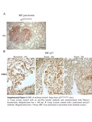
IHC • IHC refers to the process of detecting antigens in cells of a tissue section by exploiting the principle of antibodies binding specifically to antigens in biological tissues. • "immuno" refers to antibodies used in the procedure, "histo" meaning tissue and chemistry refers to the staining process. • IHC staining is widely used in the diagnosis of abnormal cells such as those found in cancerous tumors. • IHC is also widely used in basic research to understand the distribution and localization of biomarkers and differentially expressed proteins in different parts of a biological tissue. • Cytokeratins in placenta for example are confined to trophoblasts and can be used as biomarkers to detect changes occur during gestation.
Principle • Visualising an antibody-antigen interaction can be accomplished in a number of ways. • In the most common instance, an antibody is conjugated to an enzyme, such as peroxidase, that can catalyse a colour-producing reaction. • Alternatively, the antibody can be tagged to a fluorophore, such as fluorescein or rhodamine.
Sample preparation • While using the right antibodies to target the correct antigens and amplify the signal is important for visualization, complete preparation of the sample is critical to maintain cell morphology, tissue architecture and the antigenicity of target epitopes. This requires proper tissue collection, fixation and sectioning.
Procedure • Primary antibodies are raised against an antigen of interest (e.g. Cytokeratin) and are typically unconjugated (unlabelled). • Secondary antibodies are raised against Igs of the primary antibody. • The secondary antibody is usually conjugated to a linker molecule, such as biotin, that then recruits reporter molecules, or the secondary antibody is directly bound to the reporter molecule itself (fluorophore). • the most popular methods of detection are with enzyme- and fluorophore-mediated chromogenic and fluorescent detection, respectively.
Procedure • With chromogenic reporters, an enzyme label is reacted with a substrate to yield an intensely colored product that can be analyzed with an ordinary light microscope. • While the list of enzyme substrates is extensive, Alkaline phosphatase (AP) and horseradish peroxidase (HRP) are the two enzymes used most extensively as labels for protein detection. • An array of chromogenic substrates is available for use with either enzyme, including DAB or BCIP/NBT, which produce a brown or purple staining, respectively, wherever the enzymes are bound.
Procedure A B The role of the 2° biotinylated antibody in amplification the signal of detecting the CK. (A) 1° antibody provides more binding sites for the 2° antibody, so (B) more binding sites will be available for avidin/biotin complex on the surface of the 2° antibody rather than very few sites when only one antibody is used. Further amplification can be achieved if the secondary antibody is conjugated to several biotin molecules.
38-week placenta 7-week placenta
Direct method • The direct method is a one-step staining method and involves a labeled antibody (e.g. FITC-conjugated antiserum) reacting directly with the antigen in tissue sections. • While this technique utilizes only one antibody and therefore is simple and rapid, the sensitivity is lower due to little signal amplification, such as with indirect methods, and is less commonly used than indirect methods.
DMD B A Immunofluorescently stained muscle biopsy. Three muscle biopsy samples are stained fluorescent linked antibody examined fluorescent microscope. (a) Shows DMD patient with no detected dystrophin. (b) shows a manifesting carrier with positive and negative protein expression. (c) shows normal expression of dystrophin in unaffected male C