Download
1 / 35
350 likes | 374 Vues
Explore the properties of sound waves, including propagation and reflection, as well as the principles behind ultrasound imaging. Learn about the frequency and velocity of ultrasound waves and how they are used in diagnostic imaging. Discover the components of an ultrasound machine and how it generates and receives sound waves.

E N D
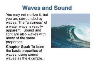
SoundWaves • Sound waves are mechanical • Require a medium for transmission • Sound is formed by the vibration or movement of air or liquid. • When a sound is made the vibrations make the surrounding particles in the air or liquid vibrate. • The sound makes the molecules compress and expand and bump into each other. • Sound waves with frequencies above the range of human hearing, i.e. 20 Hz to 20KHz.
Sound waves and ultrasound waves follow the rules of propagation and reflection similar to those that govern light waves in that they can be Reflection- to throw back the sound wave at the interface of two materials. Refraction- to bend the sound wave at the interface of two materials, i.e. the waves are transmitted. Absorption- attenuation of a sound wave by “relaxation” or frictional processes in the medium that convert acoustic energy to heat, i.e. some portion of the waves in not transmitted.
Speed Of Sound • Depends upon the compressibility of material. • γ is the speed of sound in the medium, λ is the wavelength, and f is the frequency of the wave (i.e., 1/T) γ = f λ c = f λ for X-rays • Speed is low in air, higher in soft tissue and highest in bone; air(331 m/s)< soft tissue(1540 m/s)< bone(3360 m/s) .
Ultrasound Frequency • The wavelength of ultrasound for diagnostic purposes should be on the order of 1mm or less. • The wavelength of sound decreases as frequency(MHz) increases. • Diagnostic imaging typically uses frequencies between 20 KHz to 1 MHz.
Velocity Of Ultrasound Waves • Velocity of ultrasound waves in tissue is independent of the wave frequency. • Velocity of sound is inversely proportional to compressibility . • Directly proportional to density. • Velocity in tissue is 1540 m/s • -travels 1 cm in 13 μsecs
Formation Of Ultrasound Waves • Medical ultrasound waves are generated by electrically vibrating a transducer. • - like piston in a water • The wave length of ultrasound wave is distance between two bands of compression and rarefaction.
Echolocation is the ability of certain animals to produce pulses of sound (either audible or ultrasonic) and then to receive the returning echoes which are processed by the brain to give information about obstacles.
Frequencies • Ultrasound – Greater then 20000 Hz • Infrasound – Less than 20 Hz • Therapeutic ultrasound – 0.5 to 5MHz • 1 to 3 M Hz
What is Ultrasound.. ? • Ultrasound or ultrasonography is a medical imaging technique that uses high frequency sound waves and their echoes. • An ultrasound test is a radiology technique, which uses high- frequency sound waves to produce images of the organs and structures of the body. • The technique is similar to the echolocation used by bats, whales and dolphins, as well as SONAR used by submarines.
Cont… • In ultrasound, the following events happen: • The sound waves are sent through body tissues with a device called a transducer. • The transducer is placed directly on top of the skin, which has a gel applied to the surface. • The sound waves that are sent by the transducer into the body and hit a boundary of organs • Some of the sound waves get reflected back to the probe, while some travel on further until they reach another boundary and get reflected. • The machine calculates the distance from the probe to the tissue or organ (boundaries) and the time of the each echo's return (usually on the order of millionths of a second).
Cont… • The machine displays the distances and intensities of the echoes on the screen, forming a two dimensional image. • The echo images are then recorded on a plane film and can also be recorded on videotape. • In ultrasound millions of pulses and echoes are sent and received each second. The probe can be moved along the surface of the body and angled to obtain various views. • After the ultrasound, the gel is easily wiped off. • The technical term for ultrasound testing and recording is "sonography."
Ultrasound Machine Ultrasound machine with various transducer probe
Parts of Ultrasound Machine • Transducer Probe • Central Processing Unit (CPU) • Transducer Pulse Controls • Display • Keyboard/Cursor • Disk Storage Device • Printer
Transducer Probe • It is the main part of the ultrasound machine. The transducer probe makes the sound waves and receives the echoes. It is, so to speak, the mouth and ears of the ultrasound machine. • The transducer probe generates and receives sound waves using a principle called the piezoelectric (pressure electricity) effect. • In the probe, there are one or more quartz crystals called Piezoelectric crystals. • Piezoelectric Crystal- reversible used to both excite and detect ultrasound waves.
Transducer Piezoelectric Crystals
Cont… • When an electric current is applied to these crystals, they change shape rapidly. The rapid shape changes, or vibrations, of the crystals produce sound waves. • The same crystals can be used to send and receive sound waves. • Transducer probes come in many shapes and sizes. • The shape of the probe determines its field of view, and the frequency of emitted sound waves determines how deep the sound waves penetrate and the resolution of the image.
Cont… • Transducer probes may contain one or more crystal elements. • Multiple-element probes have the advantage that is especially important for cardiac ultrasound. • Piezoelectrical transducers are used to achieve the high-frequency ultrasound energy needed for imaging and therapy. • These are suitably cut crystals, which change shape under the influence of an electric charge. • Many types of crystal can be used but the most favored are quartz, which occurs naturally, and some synthetic ceramic materials such as barium titanate and lead zirconatetitanate (PZT).
Cont…. Cont… • These crystals deform when subjected to a varying potential difference – a piezo-electric effect, is discovered by Pierre and Jacques Curie in 1880. • The principle of converting energy by applying pressure to a crystal. • The reverse of the piezoelectric effect converts the energy back to its original form. • The crystal must be cut to suitable dimensions – the most important being the thickness – so that it will resonate at the chosen frequency and so achieve maximum vibration.
Piezoelectric Effect & Ultrasound Transducers • A transducer converts one type of energy into another. • Based upon the pulse-echo principle occurring with ultrasound piezoelectric crystals, ultrasound transducers convert: • Electricity into sound = pulse • Sound into electricity = echo
Electronics Transducer • Curved Array • – crystals are placed parallel or in concentric rings • – transducer face is curved • – produces sector or pie-shaped image • Linear Array • – crystals are placed parallel • – transducer face is flat • – produces rectangular image
Electronics Transducer • Groups of piezoelectric material working singly or in groups.
Central Processing Unit • The CPU is the brain of the ultrasound machine. • The CPU is basically a computer that contains the microprocessor, memory, amplifiers and power supplies for the microprocessor and transducer probe. • The CPU sends electrical currents to the transducer probe to emit sound waves, and also receives the electrical pulses from the probes that were created from the returning echoes. • The CPU does all of the calculations involved in processing the data.
Cont… • Once the raw data are processed, the CPU forms the image on the monitor. • The CPU does all of the calculations involved in processing the data. • The CPU can also store the processed data and/or image on disk.
Transducer Pulse Control • The transducer pulse controls allow the operator, called the ultrasonographer. • To set and change the frequency and duration of the ultrasound pulses, as well as the scan mode of the machine. • The commands from the operator are translated into changing electric currents that are applied to the piezoelectric crystals in the transducer probe.
Procedure of Ultrasound • The sonographer asks the patient to take a deep breath and hold it. This brings the top of the liver out from under the ribs. • Then the transducer is either moved in a straight line in a longitudinal or transverse direction, or else rocked back and forth in one place. • Procedure will produce a two-dimensional scan on the screen. • The sonographer scans the rest of the liver and takes pictures of areas of interest
Display • The display is a computer monitor that shows the processed data from the CPU. • Displays can be black-and-white or color, depending upon the model of the ultrasound machine.
Keyboard / Cursor • Ultrasound machines have a keyboard and a cursor, such as a trackball, built in. • These devices allow the operator to add notes to and take measurements from the data.
Disk Storage • The processed data and/ or images can be stored on disk. • The disks can be hard disks, floppy disks, compact discs (CDs) or digital video discs (DVDs). • Typically, a patient's ultrasound scans are stored on a floppy disk and archived with the patient's medical records.
Printers Printer • Many ultrasound machines have thermal printers that can be used to capture a hard copy of the image from the display.